Unlock hundreds more features
Save your Quiz to the Dashboard
View and Export Results
Use AI to Create Quizzes and Analyse Results

Sign inSign in with Facebook
Sign inSign in with Google

Back Muscles Quiz: Label the Major Muscle Groups

Quick, free back anatomy quiz to test your knowledge. Instant results.

Editorial: Review CompletedCreated By: Negima SekaiUpdated Aug 28, 2025
Difficulty: Moderate
2-5mins
Learning OutcomesCheat Sheet
Paper art illustration of back muscles including trapezius, latissimus dorsi and other key groups on dark blue background

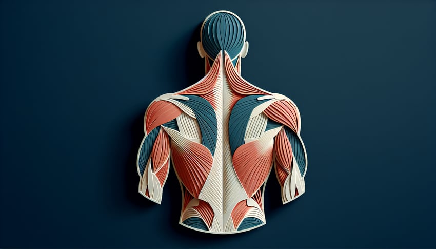
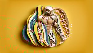
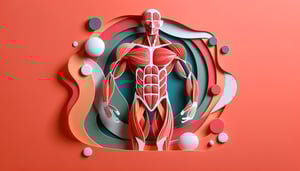
This back muscles quiz helps you label the trapezius, latissimus dorsi, rhomboids, and more on clear diagrams. Check your knowledge fast, then explore related areas with a scapula labeling quiz, a vertebrae labeling quiz, or an axial muscles quiz. Get instant feedback and keep practicing.

Which muscle primarily elevates the scapula and is innervated by the dorsal scapular nerve?
Latissimus dorsi
Levator scapulae
Serratus posterior superior
Multifidus
undefined
Which nerve provides motor innervation to the trapezius?
Spinal accessory nerve (CN XI)
Dorsal scapular nerve
Long thoracic nerve
Thoracodorsal nerve
undefined
Erector spinae is composed of iliocostalis, longissimus, and spinalis muscle groups.
True
False
undefined
Which muscle retracts the scapula and lies deep to the trapezius?
Quadratus lumborum
Rhomboid major
Rotatores
Serratus posterior inferior
undefined
The triangle of auscultation is bordered by the trapezius, latissimus dorsi, and the medial border of the scapula.
True
False
undefined
Which muscle extends and laterally flexes the vertebral column and is the most lateral column of the erector spinae?
Longissimus
Spinalis
Multifidus
Iliocostalis
undefined
The latissimus dorsi inserts on the floor of the intertubercular sulcus of the humerus.
True
False
undefined
The thoracodorsal nerve is a branch of the posterior cord of the brachial plexus and innervates latissimus dorsi.
False
True
undefined
Which muscle group is primarily responsible for contralateral rotation of the vertebral column?
Quadratus lumborum
Erector spinae (iliocostalis)
Transversospinalis (e.g., semispinalis, multifidus)
Splenius capitis
undefined
The dorsal rami of spinal nerves primarily innervate the superficial back muscles.
False
True
undefined
The rhomboid minor inserts on the medial border of the scapula at the level of the spine of the scapula.
True
False
undefined
The splenius capitis produces ipsilateral rotation of the head when acting unilaterally.
True
False
undefined
Which artery typically supplies the trapezius alongside CN XI?
Posterior intercostal artery
Transverse cervical artery (superficial branch)
Thoracodorsal artery
Dorsal scapular artery
undefined
Which structure forms the posterior layer of the thoracolumbar fascia attaching to spinous processes?
Nuchal ligament
Clavipectoral fascia
Middle scalene aponeurosis
Posterior lamina of thoracolumbar fascia
undefined
The dorsal scapular artery typically accompanies the dorsal scapular nerve deep to the levator scapulae and rhomboids.
True
False
undefined
The lumbar triangle (of Petit) is bounded by latissimus dorsi, external oblique, and the iliac crest.
True
False
undefined
The semispinalis capitis inserts onto the mastoid process of the temporal bone.
True
False
undefined
The suboccipital nerve is the dorsal ramus of C2 and innervates the suboccipital muscles.
False
True
undefined
The posterior cutaneous branches of posterior intercostal arteries contribute to the blood supply of intrinsic back muscles.
False
True
undefined
The fascia enclosing the erector spinae is continuous with the nuchal ligament superiorly and thoracolumbar fascia inferiorly.
False
True
undefined
0

Study Outcomes

  1. Identify Major Back Muscles -

    Learn to identify the trapezius, latissimus dorsi, rhomboids, and other key back muscles by name and position.

  2. Label Superficial vs Deep Layers -

    Distinguish between superficial and deep muscle layers on diagrams to enhance your understanding of back muscle anatomy.

  3. Recall Anatomical Origins and Insertions -

    Memorize the origins and insertions of each back muscle group to support accurate labeling and functional understanding.

  4. Differentiate Muscle Functions -

    Understand and explain the primary actions of each muscle, such as extension, rotation, and stabilization of the spine and shoulders.

  5. Apply Knowledge in Practical Quizzes -

    Test your anatomy skills through interactive labeling exercises, reinforcing your retention of back muscle names and functions.

Cheat Sheet

  1. Trapezius Anatomy & Function -

    In the back muscles quiz, you'll need to identify the trapezius's triangular shape spanning from the occiput to T12 and recognize its three fiber segments for scapular elevation, retraction, and depression. Use the mnemonic "Up, Back, Down" to remember upper, middle, and lower fiber actions. (Source: Gray's Anatomy, 41st ed.)

  2. Latissimus Dorsi Origins & Actions -

    The latissimus dorsi originates along the thoracolumbar fascia, iliac crest, and lower ribs and inserts into the intertubercular groove of the humerus to power shoulder extension, adduction, and internal rotation. Often called the "swimmer's muscle," it's essential in any back muscle quiz for its broad, fan-like appearance. (Source: Netter's Atlas of Human Anatomy, 7th ed.)

  3. Erector Spinae Group Breakdown -

    The erector spinae consists of iliocostalis, longissimus, and spinalis columns, critical for spinal extension and posture maintenance in back muscles labeling exercises. Remember "I Love Spaghetti" to sequence them from lateral to medial. (Source: Tortora & Nielsen, Principles of Anatomy and Physiology.)

  4. Rhomboids & Levator Scapulae Roles -

    Rhomboid major and minor retract and downwardly rotate the scapula, while levator scapulae elevates it, making these key when labeling back muscles on any musculoskeletal chart. A quick self-check: pull shoulders back to feel rhomboid engagement and shrug slightly for levator activation. (Source: American Journal of Physical Medicine & Rehabilitation.)

  5. Deep Intrinsic Stabilizers -

    Multifidus, interspinales, and rotatores sit closest to vertebrae, providing fine-tuned proprioception and segmental stability; multifidus is especially prominent in the lumbar region. Use "MRI" (Multifidus, Rotatores, Interspinales) to ace the muscles of the back quiz on deep layer identification. (Source: Journal of Orthopaedic & Sports Physical Therapy.)

Powered by: Quiz Maker