Female Reproductive System Labeling Quiz
Quick female reproductive anatomy quiz to test your knowledge. Instant feedback.
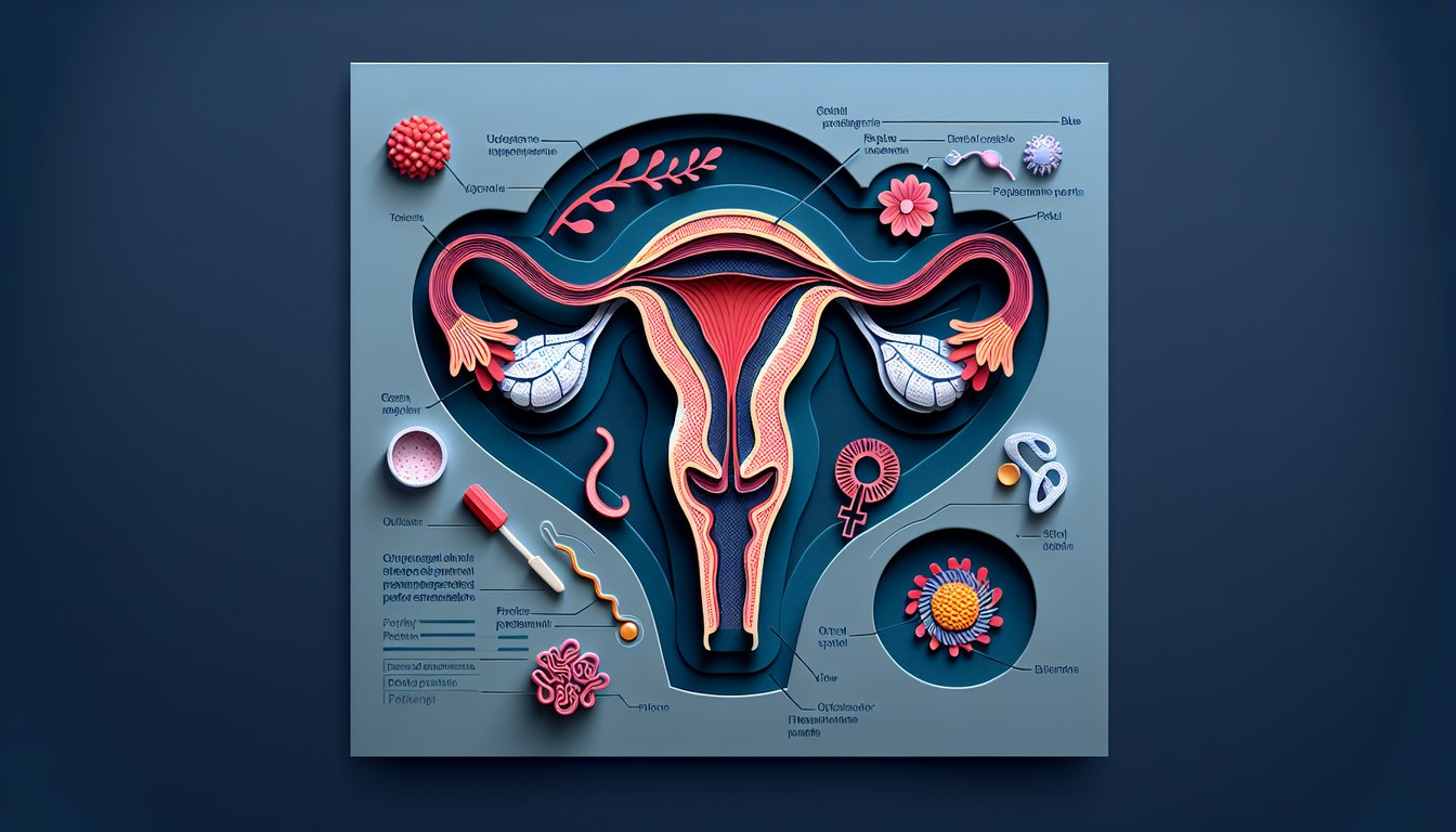
This quiz helps you label the female reproductive system and place each part, from ovaries and fallopian tubes to the uterus and cervix. To compare systems, try our male reproductive system labeling quiz or the male reproductive system quiz, and build context with a pelvic girdle labeling quiz.
Study Outcomes
- Identify Key Structures -
Recognize and name the major components of the female reproductive system, including the ovaries, fallopian tubes, uterus, and external genitalia.
- Label Reproductive Anatomy -
Accurately place labels on a detailed diagram of the female reproductive system to reinforce spatial relationships between organs.
- Describe Organ Functions -
Explain the primary roles of each labeled structure in processes such as ovulation, fertilization, and menstruation.
- Differentiate Internal vs. External Organs -
Distinguish between internal reproductive organs and external genitalia to deepen understanding of system organization.
- Explain Structure-Function Relationships -
Analyze how the anatomical features of each part contribute to overall reproductive health and function.
- Apply Anatomy Knowledge -
Use your quiz results to inform further study in biology, healthcare, or related fields by applying core reproductive anatomy concepts.
Cheat Sheet
- Ovaries -
When you label female reproductive system diagrams, start by pinpointing the almond-shaped ovaries in the pelvic cavity; they're about 3 cm long and sit on either side of the uterus. Remember "Only Fair Unicorns Crop Veggies" to recall Ovaries, Fallopian tubes, Uterus, Cervix, Vagina in order. The ovaries release oocytes and secrete estrogen and progesterone, driving the menstrual cycle (NIH).
- Fallopian Tubes -
Divide each tube into infundibulum (with fimbriae), ampulla (fertilization zone), isthmus, and intramural segment; use "I Am Ingesting Irish Stew" to memorize. Fimbriae sweep the oocyte into the tube, while peristalsis and cilia guide it toward the uterus (American College of Obstetricians and Gynecologists). Knowing these subdivisions helps you ace any female reproductive anatomy quiz.
- Uterus -
Identify the pear-shaped uterus and its three layers - perimetrium (outer), myometrium (muscular), and endometrium (inner); use the acronym "P.M.E." to recall them. The endometrium builds up and sheds during the menstrual cycle, which you can diagram with a simple 28-day timeline (e.g., days 1 - 14 follicular, day 14 ovulation, days 15 - 28 luteal). Mastering these layers boosts confidence in labeling the female reproductive system.
- Cervix -
Locate the cervix as the narrow, cylindrical neck of the uterus with two openings: the internal and external os. Note the transformation zone (squamocolumnar junction) where sampling occurs in Pap smears - this landmark is vital for accurate diagrams (World Health Organization). Observing how cervical mucus changes during ovulation also deepens your anatomical insight.
- Vagina and Vestibular Glands -
Label the vagina as the fibromuscular canal extending from the cervix to the vulva, and mark its transverse rugae which allow expansion. Don't forget Bartholin's glands (located at 5 and 7 o'clock) that secrete lubrication - imagine two "Bartho-ling lights" at the vestibule. Recognizing these features enhances your skills in any female reproductive diagram quiz.