Unlock hundreds more features
Save your Quiz to the Dashboard
View and Export Results
Use AI to Create Quizzes and Analyse Results

Sign inSign in with Facebook
Sign inSign in with Google

Take the Nervous System Quiz Now!

Dive into our neurological system quiz and boost your CNS know-how!

Difficulty: Moderate
2-5mins
Learning OutcomesCheat Sheet
Paper art illustration for a nervous system quiz on a dark blue background

This Nervous System Quiz helps you review how neurons send signals, how the brain and spinal cord work, and where reflexes start. Use it to spot gaps before an exam. For more practice, try the extended practice quiz or the neurology version .

What is the basic functional unit of the nervous system?
Axon
Neuron
Dendrite
Glial cell
Neurons are specialized cells that transmit electrical and chemical signals throughout the body, forming the nervous system's basic unit. Each neuron consists of dendrites, a cell body, and an axon that work together to process and convey information. Glial cells support neurons but do not conduct impulses. Learn more at .
Which two divisions make up the peripheral nervous system?
Central and Peripheral
Sensory and Motor
Somatic and Autonomic
Sympathetic and Parasympathetic
The peripheral nervous system is divided into the somatic division, which controls voluntary movements, and the autonomic division, which regulates involuntary functions. The autonomic division further splits into sympathetic and parasympathetic branches. Central and peripheral describe the two overarching nervous system groups, not subdivisions of the PNS. More details at .
What is the primary function of the myelin sheath?
Store neurotransmitters
Increase the speed of electrical conduction
Generate new neurons
Anchor neurons in place
Myelin is a fatty insulating layer that surrounds axons and allows electrical impulses to travel quickly and efficiently along the nerve fiber. By preventing ion leakage, it increases conduction velocity through saltatory conduction. It does not store neurotransmitters or generate neurons. Read more at .
Approximately what is the resting membrane potential of a typical neuron?
+40 mV
-100 mV
-70 mV
0 mV
A typical neuron at rest has a membrane potential of around -70 millivolts, with the inside more negative than the outside. This electrochemical gradient is maintained by ion pumps and channels, notably the sodium-potassium ATPase. Deviations from this value can affect neuronal excitability. See .
Which neurotransmitter is released at the neuromuscular junction to stimulate muscle contraction?
GABA
Acetylcholine
Dopamine
Serotonin
Acetylcholine is the neurotransmitter that motor neurons release at the neuromuscular junction to trigger muscle fiber contraction. It binds to nicotinic receptors on the muscle cell membrane, causing depolarization. Dopamine, GABA, and serotonin are primarily used within the central nervous system. More information at .
Which structures comprise the central nervous system?
Brain and spinal cord
Cranial nerves and spinal nerves
Spinal cord and peripheral nerves
Brain and peripheral nerves
The central nervous system (CNS) consists of the brain and spinal cord, which process information and coordinate activity throughout the body. Peripheral nerves lie outside the CNS and form the peripheral nervous system. Cranial and spinal nerves are part of the peripheral division. Read more at .
What is the role of oligodendrocytes in the central nervous system?
Act as phagocytes
Produce cerebrospinal fluid
Form myelin sheaths around axons
Maintain the blood-brain barrier
Oligodendrocytes are glial cells in the CNS that wrap around axons to form myelin sheaths, which increase action potential conduction speed. Microglia act as phagocytes, ependymal cells produce cerebrospinal fluid, and astrocytes help maintain the blood-brain barrier. More at .
What is the primary function of the nodes of Ranvier?
Anchor dendrites
Release neurotransmitters
Generate new neurons
Enable rapid saltatory conduction
Nodes of Ranvier are gaps in the myelin sheath along an axon where voltage-gated sodium channels are concentrated. They allow the action potential to 'jump' from node to node in saltatory conduction, greatly increasing conduction velocity. They do not release neurotransmitters or generate neurons. Learn more at .
What best describes saltatory conduction in a myelinated axon?
The impulse travels continuously along the entire axon
The impulse spreads across multiple dendrites
The impulse jumps from one node of Ranvier to the next
The impulse crosses the synaptic cleft
Saltatory conduction refers to the rapid propagation of action potentials along myelinated axons by 'jumping' between nodes of Ranvier. This mechanism increases conduction speed and efficiency. Unmyelinated axons conduct impulses continuously rather than in leaps. More at .
Which brain region is primarily responsible for coordinating balance and fine motor control?
Cerebellum
Thalamus
Medulla oblongata
Hypothalamus
The cerebellum integrates sensory input with motor commands to coordinate balance, posture, and fine motor movements. It receives proprioceptive information from muscles and joints and adjusts motor output accordingly. The thalamus acts as a relay station, the medulla controls autonomic functions, and the hypothalamus regulates homeostasis. Details at .
Which neurotransmitter is predominantly used by postganglionic sympathetic neurons?
GABA
Acetylcholine
Norepinephrine
Dopamine
Postganglionic sympathetic neurons primarily release norepinephrine at their synapses to activate adrenergic receptors on target organs. Acetylcholine is used by preganglionic sympathetic and parasympathetic neurons, while dopamine and GABA serve different functions in the CNS. More at .
Which system within the brain is most associated with emotion and memory?
Limbic system
Reticular formation
Corpus callosum
Basal ganglia
The limbic system, including structures like the hippocampus and amygdala, plays a central role in processing emotions and forming memories. The basal ganglia regulate movement, the reticular formation manages arousal, and the corpus callosum connects the cerebral hemispheres. Learn more at .
What cognitive functions are most commonly lateralized to the left cerebral hemisphere?
Language and analytical reasoning
Autonomic regulation
Emotional processing
Spatial orientation and face recognition
In most individuals, the left hemisphere specializes in language processing, mathematical reasoning, and analytical tasks. The right hemisphere more often handles spatial awareness and face recognition. Emotional processing and autonomic functions involve multiple brain areas. More at .
Which structure connects the left and right cerebral hemispheres?
Fornix
Corpus callosum
Hippocampus
Thalamus
The corpus callosum is a thick bundle of nerve fibers that links the two cerebral hemispheres, allowing communication between them. The fornix connects hippocampal structures, the thalamus is a relay center, and the hippocampus is involved in memory formation. Learn more at .
What is the correct order of elements in a simple reflex arc?
Effector ? Motor neuron ? Interneuron ? Receptor
Sensory neuron ? Motor neuron ? Receptor ? Effector
Motor neuron ? Interneuron ? Sensory neuron ? Effector
Receptor ? Sensory neuron ? Interneuron ? Motor neuron
A reflex arc begins at a receptor that detects a stimulus, sends a signal via a sensory neuron to an interneuron in the spinal cord, which activates a motor neuron that triggers the effector organ. This pathway allows rapid, involuntary responses. Other sequences listed are incorrect. More at .
What cells and structures primarily form the blood-brain barrier?
Microglia and pericytes
Endothelial cells with tight junctions and astrocyte endfeet
Schwann cells and basal lamina
Oligodendrocytes and myelin
The blood-brain barrier is created by endothelial cells joined by tight junctions, supported by astrocyte endfeet and pericytes, which regulate molecule passage into the CNS. Oligodendrocytes form myelin, microglia act as immune cells, and Schwann cells myelinate peripheral nerves. More details at .
Which cortical area is primarily involved in planning voluntary motor movements?
Premotor cortex
Primary motor cortex
Prefrontal cortex
Primary somatosensory cortex
The premotor cortex, located anterior to the primary motor cortex, is critical for planning and selecting voluntary movements. The primary motor cortex executes movement commands, while the prefrontal cortex is involved in decision-making. The somatosensory cortex processes sensory input. Read more at .
What distinguishes temporal summation from spatial summation at a neuronal synapse?
Temporal summation only occurs in muscle cells, spatial only in neurons
Temporal summation involves successive signals over time at one synapse, spatial summation involves simultaneous signals at multiple synapses
They are identical processes under different names
Temporal summation is inhibitory, spatial summation is excitatory
Temporal summation is the additive effect of multiple action potentials arriving in quick succession at the same synapse, whereas spatial summation combines simultaneous inputs from different synapses on the same neuron. Both processes influence whether the postsynaptic membrane reaches threshold. They are distinct and essential for neuronal integration. More at .
Which spinal nerve roots carry sensory information into the spinal cord?
Anterior roots
Dorsal roots
Ventral roots
Lateral roots
Dorsal (posterior) roots of spinal nerves contain afferent fibers that carry sensory information from the periphery into the spinal cord. Ventral (anterior) roots contain efferent motor fibers. There are no separate lateral or anterior roots in spinal nerve anatomy. More at .
What is the role of lateral inhibition in sensory pathways?
Enhance contrast between stimulated and neighboring areas
Transmit pain signals to the brain
Increase overall signal intensity
Prevent overstimulation of receptors
Lateral inhibition is a neural mechanism where activated neurons suppress the activity of neighboring neurons, sharpening the contrast and improving sensory resolution. It is important in processes like edge detection in vision. It does not increase overall signal intensity or specifically transmit pain. Learn more at .
What is the main function of the substantia nigra in the basal ganglia circuit?
Produce dopamine to regulate movement
Form long-term memories
Relay sensory information to the cortex
Coordinate visual reflexes
The substantia nigra pars compacta produces dopamine, which modulates basal ganglia pathways critical for the initiation and control of movement. Degeneration of these neurons causes Parkinson's disease. It is not primarily involved in sensory relay, memory formation, or visual reflexes. See .
How do G protein-coupled receptors (GPCRs) typically transduce extracellular signals in neurons?
Directly open ion channels without intermediates
Activate intracellular second messengers via G proteins
Phagocytose extracellular molecules
Enter the nucleus to alter gene transcription
GPCRs activate heterotrimeric G proteins upon ligand binding, which then influence intracellular second messengers like cAMP or IP3 to propagate the signal. They do not directly open ion channels, enter the nucleus, or phagocytose molecules. Learn more at .
What key role does calcium (Ca²?) play in synaptic transmission?
Blocks sodium channels to end the action potential
Degrades neurotransmitters in the synaptic cleft
Triggers synaptic vesicle fusion and neurotransmitter release
Forms tight junctions in the blood-brain barrier
When an action potential reaches the synaptic terminal, voltage-gated calcium channels open, allowing Ca²? influx. The rise in intracellular calcium triggers synaptic vesicles to fuse with the presynaptic membrane and release neurotransmitters. Calcium does not block sodium channels or degrade neurotransmitters. More at .
Which variables are included in the Nernst equation to calculate the equilibrium potential for a single ion?
Action potential amplitude and duration
Synaptic cleft width and neurotransmitter affinity
Membrane capacitance and resistance
Ion concentration ratio, temperature, gas constant, Faraday's constant, and ion valence
The Nernst equation calculates an ion's equilibrium potential based on the ratio of its extracellular to intracellular concentration, the absolute temperature, the universal gas constant, Faraday's constant, and the ion's valence. This potential is the voltage at which there is no net ion flow across the membrane. It does not involve membrane capacitance or synaptic factors. See .
Which sequence of events best describes the early mechanism of long-term potentiation (LTP) at glutamatergic synapses?
Metabotropic receptor inhibition, reduced cAMP, decreased neurotransmitter release
NMDA receptor activation, Ca²? influx, activation of CaMKII, insertion of AMPA receptors
Voltage-gated Na? channel opening, action potential backpropagation, K? efflux
GABA release, Cl? influx, hyperpolarization, AMPA removal
During early LTP at excitatory synapses, strong stimulation relieves the Mg²? block on NMDA receptors, allowing Ca²? influx. The calcium activates kinases such as CaMKII, which promote insertion and phosphorylation of AMPA receptors, strengthening synaptic transmission. This process is critical for learning and memory. More details at .
0
{"name":"What is the basic functional unit of the nervous system?", "url":"https://www.quiz-maker.com/QPREVIEW","txt":"What is the basic functional unit of the nervous system?, Which two divisions make up the peripheral nervous system?, What is the primary function of the myelin sheath?","img":"https://www.quiz-maker.com/3012/images/ogquiz.png"}

Study Outcomes

  1. Identify Nervous System Structures -

    Recognize and name the key components of both the central and peripheral nervous systems, including major brain regions, neurons, and the spinal cord.

  2. Explain Neuron Function -

    Describe the anatomy of a neuron and explain how electrical impulses and neurotransmitters facilitate nerve signal transmission.

  3. Analyze Neural Pathways -

    Trace the flow of sensory and motor signals through neural circuits to understand how the body processes and responds to stimuli.

  4. Differentiate CNS and PNS Roles -

    Compare the functions of the central nervous system versus the peripheral nervous system and their contributions to overall neurological health.

  5. Apply Knowledge in Quiz Scenarios -

    Use your understanding of nervous system anatomy and physiology to tackle varied quiz questions and improve retention for future exams.

Cheat Sheet

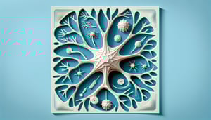
  1. Neuron Structure & Signal Propagation -

    Review the parts of a neuron - dendrites, soma, axon and myelin sheath - and how an action potential travels via saltatory conduction between Nodes of Ranvier. A handy mnemonic "D.A.S.A." (Dendrite→Axon→Soma→Axon terminal) helps remember signal flow. Multiple sclerosis (MS) illustrates demyelination slowing conduction (NIH NINDS).

  2. Synaptic Transmission & Key Neurotransmitters -

    Understand chemical synapses: Ca²❺ influx triggers vesicle fusion via SNARE proteins, releasing neurotransmitters into the cleft. Remember acetylcholine at the neuromuscular junction and dopamine in reward pathways (Purves et al., Neuroscience). In a neurological system quiz, linking transmitter to function boosts recall - "Dopamine = Drive."

  3. Central vs. Peripheral Divisions -

    Distinguish the CNS (brain and spinal cord) from the PNS (cranial/spinal nerves and ganglia) as defined by the American Physiological Society. Use the mnemonic "SAME DAVE" to recall sensory afferents (SAME) and motor efferents (DAVE). This core concept often forms the basis of a quiz about the nervous system question on pathway organization.

  4. Brain Region Functions & Mnemonics -

    Map the prosencephalon, mesencephalon and rhombencephalon to forebrain, midbrain and hindbrain roles in cognition, vision and balance (University of Michigan). Try "TeDiMeMy" (Telencephalon, Diencephalon, Mesencephalon, Metencephalon, Myelencephalon) to list subregions. This tip can make your neuron system quiz swift and accurate.

  5. Spinal Reflex Arcs & Clinical Significance -

    Master the monosynaptic stretch reflex (e.g., patellar reflex) and the polysynaptic withdrawal reflex for rapid protective responses. Trace each arc: receptor→sensory neuron→(interneuron)→motor neuron→effector, which you'll see on most central nervous system tests. Clinically, reflex grading (0 - 4+) reveals lesion localization (NIH).

Powered by: Quiz Maker