Ear labeling quiz: name parts on the ear diagram
Quick, free ear diagram quiz to test your knowledge. Instant results.
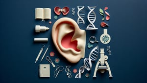
This ear labeling quiz helps you identify and name each part on a clear ear diagram. Practice at your own pace, check what you know, and see results as you go. When you want more, try our parts of the ear quiz or dive deeper with an inner ear anatomy quiz.
Study Outcomes
- Understand Ear Labeling Basics -
Learn to distinguish the outer, middle, and inner ear regions to build a solid foundation for ear labeling.
- Identify Key Structures -
Practice identifying critical components on a diagram of the ear with labels, such as the cochlea, ossicles, and auditory canal.
- Recall Functional Roles -
Memorize each ear part's function to link anatomy with physiological roles in hearing and balance.
- Apply Labeling Techniques -
Use ear anatomy labeling strategies to accurately label the ear in different diagrams and learning materials.
- Analyze Common Errors -
Examine frequent mistakes in ear labeling to improve precision when you label the ear during the quiz.
- Evaluate Your Proficiency -
Assess your performance with instant feedback in the interactive ear anatomy quiz to track progress and confidence.
Cheat Sheet
- Divisions of the Ear -
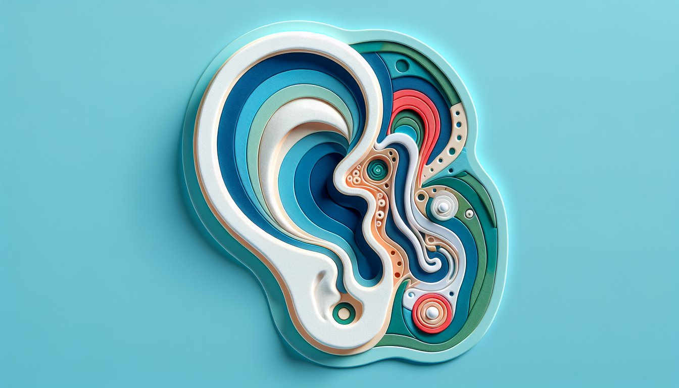
Understanding the three main regions - outer, middle, and inner ear - is foundational for any ear anatomy labeling exercise. Remember the mnemonic "O-M-I" (Outer, Middle, Inner) to keep the sequence straight when you label the diagram of the ear with labels. This tripartite division is detailed in resources like NIH's MedlinePlus, ensuring you have a reliable framework before you start your ear labeling quiz.
- Outer Ear Components -
Key landmarks include the pinna (auricle), external auditory canal, and the tympanic membrane (eardrum). Use the acronym "PAT" (Pinna, Auditory canal, Tympanic membrane) to quickly recall these parts when you label the ear. Academic sources such as Gray's Anatomy Online provide high-resolution diagrams that are perfect for practicing ear anatomy labeling.
- Middle Ear Ossicles -
The malleus, incus, and stapes form a lever system that transmits sound; a popular mnemonic is "M-I-S" (Malleus, Incus, Stapes) in lateral-to-medial order. Each tiny bone amplifies vibrations - refer to the American Academy of Otolaryngology for detailed morphology and function. When you label the ear diagram, ensure you correctly place these ossicles in the middle ear cavity.
- Inner Ear Structures -
The cochlea, vestibule, and semicircular canals are critical for hearing and balance; picture a "CSV" spreadsheet - CoClea, Semicircular canals, Vestibule - to lock these in memory. The spiral-shaped cochlea houses hair cells that transduce sound waves into electrical signals (see research from the Journal of Neuroscience). Accurate ear labeling demands precise identification of these fluid-filled chambers on your ear anatomy quiz.
- Eustachian Tube Function -
Connecting the middle ear to the nasopharynx, the Eustachian tube equalizes air pressure and drains fluids, a fact often tested in ear anatomy labeling sections. Recall the phrase "Equalize or Compromise" to remember why this tube is vital during pressure changes (e.g., flying). Official guidelines from the AAO-HNS highlight its role, so be sure to label it correctly when you take the interactive ear labeling quiz.