Heart Anatomy Quiz: Stroke Volume, Chambers, and Valves
Quick, free cardiac anatomy quiz with stroke volume questions. Instant results.

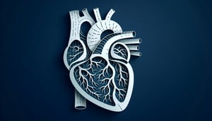
This heart anatomy quiz helps you check your understanding of chambers, valves, and stroke volume, with instant feedback. Use it to spot weak areas before class or exams. If you want to focus on structure and labeling, try the parts of the heart quiz. To broaden your view of circulation and pathways, follow up with the blood vessels quiz.
Study Outcomes
- Identify Cardiac Structures -
Learners will be able to recognize and name the major components of the heart, including chambers, valves, and vessels, through targeted heart anatomy quiz questions.
- Describe Chamber Functions -
Participants will understand the roles of the atria and ventricles in blood flow and how each chamber contributes to overall heart function.
- Trace Blood Circulation Pathways -
Users will be able to map the journey of blood through the heart and into the cardiovascular system, reinforcing their knowledge in a cardiovascular system quiz format.
- Recall Heart Trivia -
Quiz-takers will improve their retention of key heart facts and intriguing cardiovascular system trivia to boost confidence in heart trivia questions.
- Evaluate Heart Function Knowledge -
Participants will assess and reinforce their understanding of heart physiology by completing multiple-choice challenges in the heart function test.
Cheat Sheet
- Cardiac Blood Flow Pathway -
Review the sequential route of blood: right atrium → right ventricle → pulmonary arteries → lungs → pulmonary veins → left atrium → left ventricle → aorta, as outlined by the American Heart Association. Use the mnemonic "Try Pulling My Leg At Vacation" to lock in this order for heart trivia questions and cardiovascular system quiz prep. Understanding this flow is crucial for any heart anatomy quiz or heart function test.
- Chamber Pressure and Function -
Distinguish atria from ventricles by pressure and wall thickness: atria generate low pressure to fill ventricles, while ventricles produce high pressure to eject blood, according to Gray's Anatomy. Remember that left ventricular pressure exceeds right ventricular pressure due to systemic circulation resistance, a common heart quiz topic. Relating structure to function helps you ace both heart anatomy quiz and cardiovascular system quiz items.
- Phases of the Cardiac Cycle -
Master systole (contraction) and diastole (relaxation) phases and link them to heart sounds S1 and S2; S1 marks AV valve closure, S2 marks semilunar valve closure, per the American Physiological Society. Visualize pressure - volume loops or use the simple phrase "Lub-Dub = Systole-Diastole" for heart trivia questions. This understanding underpins many heart function test questions on ejection fraction and stroke volume.
- Electrical Conduction System -
Learn the impulse pathway: SA node → AV node → bundle of His → Purkinje fibers, as described in the Circulation Journal. Use "Some Adventurers Buy Pie" to recall each component for your next heart anatomy quiz or cardiovascular system quiz. Recognizing conduction order is vital for understanding arrhythmias on an ECG in a heart quiz scenario.
- Cardiac Output Formula -
Memorize the core formula CO = HR × SV (cardiac output equals heart rate times stroke volume), with a normal range of 4 - 8 L/min per NIH guidelines. Practice calculations by adjusting HR or SV to see how stress, exercise, or pathology impacts output in heart function test questions. This formula is frequently tested in both heart trivia questions and professional cardiovascular system quiz settings.