Unlock hundreds more features
Save your Quiz to the Dashboard
View and Export Results
Use AI to Create Quizzes and Analyse Results

Sign inSign in with Facebook
Sign inSign in with Google

Thorax Anatomy Quiz: Can You Ace Your Chest Anatomy?

Think You Can Ace the Chest Anatomy Quiz?

Difficulty: Moderate
2-5mins
Learning OutcomesCheat Sheet
Paper art illustration for thorax anatomy quiz on golden yellow background

This thorax anatomy quiz helps you practice chest anatomy by naming bones, muscles, and organs across 20 questions, including sternum, ribs, and organ placement. Get instant feedback to spot gaps before an exam or lab. Start the quiz and see where you stand.

Which bone articulates with the medial ends of the clavicles?
Scapula
Atlas
Humerus
Manubrium of the sternum
The clavicles articulate medially with the manubrium, which is the upper portion of the sternum. This articulation forms the sternoclavicular joint and is unique to the manubrium. The scapula and humerus articulate with the lateral end of the clavicle, while the atlas is part of the cervical spine.
What is the central part of the sternum called?
Xiphoid process
Jugular notch
Body of the sternum
Manubrium
The sternum is composed of the manubrium, the body, and the xiphoid process. The body is the longest and central portion that lies between the manubrium above and the xiphoid process below. The jugular notch is a feature of the manubrium.
How many pairs of ribs are normally present in the human thorax?
11
12
10
13
There are typically 12 pairs of ribs in the human thorax, each articulating posteriorly with the thoracic vertebrae. The first seven are true ribs, the next three are false ribs, and the last two are floating ribs. This arrangement provides both protection and flexibility to the thoracic cage.
Which ribs are classified as floating ribs?
11th and 12th ribs
7th and 8th ribs
5th and 6th ribs
9th and 10th ribs
Floating ribs are the 11th and 12th pairs because they have no anterior attachment to the sternum or costal cartilage. They articulate only with the vertebrae in the back. Their free anterior ends allow for a greater range of movement.
What type of cartilage connects the true ribs to the sternum?
Articular cartilage
Hyaline cartilage
Fibrocartilage
Elastic cartilage
The costal cartilages that directly connect the true ribs (ribs 1 - 7) to the sternum are composed of hyaline cartilage. This type of cartilage provides both strength and some flexibility to the anterior thoracic wall. Fibrocartilage and elastic cartilage are found in other parts of the body.
Which muscle separates the thoracic cavity from the abdominal cavity?
Transversus abdominis
External oblique
Diaphragm
Serratus anterior
The diaphragm is a dome-shaped musculotendinous partition that separates the thoracic cavity from the abdominal cavity. It is the primary muscle of respiration and changes shape during breathing cycles. No other muscle spans this entire boundary region.
Which muscle is primarily responsible for depressing the ribs during forced expiration?
Serratus anterior
External intercostals
Serratus posterior superior
Internal intercostals
The internal intercostal muscles run obliquely from the rib above to the rib below and act to depress the ribs during forced expiration. In contrast, the external intercostals elevate ribs during inspiration. Serratus muscles have different attachments and functions.
What is the name of the cartilage that links the anterior ends of the true ribs to the sternum?
Elastic cartilage
Costal cartilage
Fibrocartilage
Articular cartilage
Costal cartilages are bars of hyaline cartilage that connect the anterior ends of ribs 1 - 7 (true ribs) to the sternum. They allow flexibility and expansion of the thoracic cage during respiration. They are not made of fibrocartilage or elastic cartilage.
Which vein primarily drains blood from the anterior thoracic wall?
Intercostal vein
Hemiazygos vein
Azygous vein
Internal thoracic vein
The internal thoracic (mammary) vein runs parallel to the sternum and drains the anterior thoracic wall before emptying into the brachiocephalic vein. The azygous and hemiazygous systems drain the posterior thoracic wall, while intercostal veins are tributaries.
Which nerve provides motor innervation to the diaphragm?
Thoracic sympathetic trunk
Phrenic nerve
Vagus nerve
Intercostal nerves
The phrenic nerve, arising from cervical roots C3 - C5, provides the sole motor supply to the diaphragm. Without phrenic nerve function, the diaphragm cannot contract effectively. The vagus nerve carries parasympathetic fibers but does not motor-innervate the diaphragm.
Which pleural layer directly covers the lung surface?
Parietal pleura
Mediastinal pleura
Visceral pleura
Costal pleura
The visceral pleura is the serous membrane that adheres tightly to the lung surface, reflecting into the fissures. The parietal pleura lines the thoracic cavity walls. Different parts of parietal pleura are named by region (costal, mediastinal, diaphragmatic).
Which muscle lies between the ribs and assists in elevation of the rib cage during inspiration?
External intercostals
Subcostalis
Pectoralis minor
Serratus anterior
External intercostal muscles run obliquely between adjacent ribs, elevating the ribs and increasing the transverse dimension of the thoracic cavity during inspiration. Internal and innermost intercostals function in forced expiration.
The sternal angle (Angle of Louis) corresponds with which vertebral level or intervertebral disc?
T2 - T3
T5 - T6
T4 - T5
T3 - T4
The sternal angle at the junction of the manubrium and body of the sternum lies opposite the T4 - T5 intervertebral disc. It serves as an important clinical landmark for rib counting and mediastinal divisions.
Which artery branches directly from the subclavian artery to supply the anterior thoracic wall?
Superior epigastric artery
Posterior intercostal artery
Lateral thoracic artery
Internal thoracic artery
The internal thoracic artery arises from the subclavian artery and descends parallel to the sternum, giving off anterior intercostal branches to the thoracic wall. The posterior intercostals come from the thoracic aorta. The superior epigastric is a terminal branch of the internal thoracic.
Compared to the left main bronchus, the right main bronchus is generally:
More vertical and longer
Narrower and longer
Wider and shorter
More horizontal and longer
The right main bronchus is wider, shorter, and more vertical than the left, making it a common site for aspirated objects. The left bronchus is narrower, longer, and more horizontal due to the position of the heart.
The thoracic duct typically drains lymph into which venous structure?
Left brachiocephalic vein
Right subclavian vein
Right internal jugular vein
Left subclavian vein
The thoracic duct empties into the venous angle formed by the junction of the left subclavian and left internal jugular veins, which is anatomically the left brachiocephalic vein. This allows lymph from most of the body to rejoin the bloodstream.
Which small muscle originates on the inner surface of one rib and inserts on the inner surface of a rib two levels below?
Subcostalis
Innermost intercostal
Internal intercostal
External intercostal
The subcostalis muscles lie on the inner surface of the posterior thoracic wall, spanning one or two ribs to insert on the inner surface of a rib below. They assist with depressing the ribs. Innermost intercostals occupy a different layer, and the external and internal intercostals run between adjacent ribs only.
In the costal groove of a rib, structures pass in which order from superior to inferior?
Nerve, Artery, Vein
Artery, Vein, Nerve
Vein, Nerve, Artery
Vein, Artery, Nerve
The mnemonic VAN (Vein, Artery, Nerve) describes the arrangement of intercostal vessels and nerve in the costal groove from superior to inferior. This is important for clinical procedures to avoid neurovascular damage.
Which subdivision of the mediastinum contains the thymus in adults?
Superior mediastinum
Posterior mediastinum
Anterior mediastinum
Middle mediastinum
In adults, the remnant of the thymus resides in the anterior (inferior) mediastinum, just posterior to the sternum. The superior mediastinum sits above the pericardium, while the middle contains the heart. The posterior mediastinum lies behind the pericardium.
What anatomical landmark marks the inferior boundary of the thoracic cage?
Infrasternal (subcostal) angle
Transpyloric plane
Costal margin
Xiphoid process
The infrasternal or subcostal angle is formed by the diverging costal margins and marks the inferior boundary of the thoracic cage. It lies at the level of the 7th costal cartilages. The xiphoid process is just above this angle.
How many thoracic vertebrae are typically found in the human spine?
9
12
5
7
The typical adult spine contains 12 thoracic vertebrae, each articulating with a pair of ribs. This arrangement provides structural support for the thoracic cage. There are 7 cervical and 5 lumbar vertebrae in a standard vertebral column.
Why is the sternal angle clinically significant?
It divides the superior and inferior mediastinum
It corresponds to the T4 - T5 vertebral level
All of the above
It marks the level of the second costal cartilage
The sternal angle is important because it marks the articulation of the second ribs, corresponds to the T4 - T5 intervertebral disc, and demarcates the boundary between the superior and inferior mediastinum. It is a key surface landmark in thoracic anatomy.
Pain from the parietal pleura is conveyed by which nerves?
Phrenic nerve only
Phrenic and intercostal nerves
Vagus nerves
Sympathetic trunks
Sensitivity to pain from the parietal pleura is carried by both the intercostal nerves (costal and peripheral diaphragmatic pleura) and the phrenic nerve (mediastinal and central diaphragmatic pleura). The vagus has visceral sensory fibers but not pain fibers for parietal pleura.
What is the name of the recess where pleural fluid commonly accumulates at the lung base when upright?
Cardiac notch
Pulmonary sinus
Costomediastinal recess
Costodiaphragmatic recess
The costodiaphragmatic recess lies between the diaphragm and the chest wall at the lung base. It is the deepest pleural recess and the first place pleural fluid accumulates in an upright patient. The costomediastinal recess lies anteriorly, while the cardiac notch is an indentation on the left lung.
During thoracentesis, where is the safest location to insert the needle to avoid injury to the intercostal neurovascular bundle?
Just above the superior border of the lower rib
Just below the inferior border of the upper rib
Directly in the costal groove
At the midpoint of the intercostal space
The intercostal neurovascular bundle runs along the inferior margin of each rib in the costal groove, so the safest approach is just above the superior border of the lower rib to avoid these structures. This technique minimizes the risk of vascular or nerve injury during thoracentesis.
0
{"name":"Which bone articulates with the medial ends of the clavicles?", "url":"https://www.quiz-maker.com/QPREVIEW","txt":"Which bone articulates with the medial ends of the clavicles?, What is the central part of the sternum called?, How many pairs of ribs are normally present in the human thorax?","img":"https://www.quiz-maker.com/3012/images/ogquiz.png"}

Study Outcomes

  1. Identify Thoracic Bones -

    Understand how to recognize and name the ribs, sternum, and thoracic vertebrae that form the structural framework of the chest cavity.

  2. Locate Chest Muscles -

    Learn to pinpoint major muscles within the thorax, including the diaphragm and intercostal muscles, and describe their roles in respiration.

  3. Recognize Thoracic Organs -

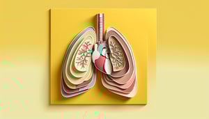
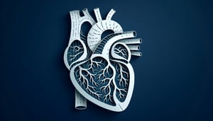
    Be able to identify the position and basic functions of vital organs such as the heart, lungs, and trachea within the human thorax.

  4. Analyze Structural Relationships -

    Examine how bones, muscles, and organs interact within the chest cavity to support breathing and protect internal structures.

  5. Apply Knowledge in Quiz Format -

    Use your understanding of chest anatomy to confidently answer multiple-choice questions in the anatomy thorax quiz and chest anatomy quiz.

Cheat Sheet

  1. Bony Thorax Composition -

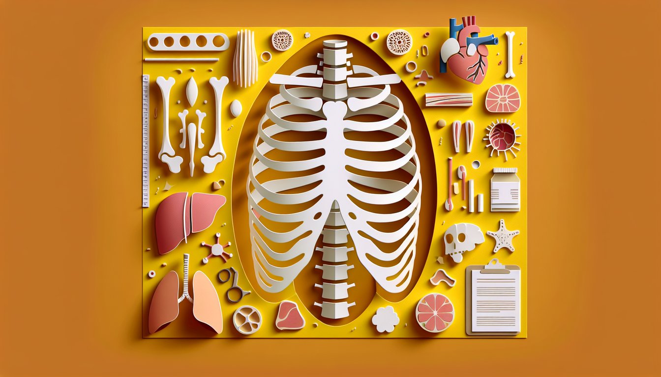
    The bony thorax consists of 12 paired ribs, costal cartilages, and the sternum, providing protection and support for thoracic organs. According to Gray's Anatomy (42nd ed.), ribs 1 - 7 are true, 8 - 10 are false, and 11 - 12 are floating - mnemonic "7 Up, 5 Down, 2 Floating." Recognizing these distinctions will help you excel on the anatomy thorax quiz.

  2. Intercostal Spaces and Neurovascular Bundle -

    Intercostal spaces contain the neurovascular bundle - vein, artery, nerve (VAN) in that order along the costal groove. A popular mnemonic is "VAN in the VANderwall" to recall Vein-Artery-Nerve from superior to inferior. This neurovascular arrangement is a staple in any thorax anatomy test and chest cavity quiz.

  3. Diaphragmatic Hiatuses -

    The diaphragm features three major hiatuses: Caval (T8) for the IVC, Esophageal (T10) for the esophagus and vagus nerves, and Aortic (T12) for the aorta and thoracic duct. Use the mnemonic "I 8 10 Eggs At 12" to memorize these openings efficiently. Mastering diaphragmatic anatomy is essential for your chest anatomy quiz and clinical correlations.

  4. Mediastinal Compartments -

    The mediastinum divides into superior and inferior regions (further split into anterior, middle, and posterior inferior compartments) with distinct organs in each. Superior houses the thymus and great vessels; middle holds the heart and pericardium; posterior contains the esophagus and descending aorta, as described in medical school texts. Understanding these compartments is critical for the human thorax quiz and thorax anatomy test.

  5. Pulmonary Anatomy and Pleura -

    The lungs are asymmetrical: the right has three lobes (via horizontal and oblique fissures) and the left has two lobes plus a lingula, per American Thoracic Society guidelines. The visceral and parietal pleura form separate cavities that reduce friction during respiration - an important clinical concept. Remember "Right 3, Left 2 plus Lingula" for quick recall on your chest anatomy quiz.

Powered by: Quiz Maker