Knee ligaments quiz: test your knee anatomy knowledge
Quick, free knee anatomy quiz. Instant results and helpful review.
This knee anatomy quiz helps you spot key knee ligaments, the menisci, cartilage, and bony landmarks, and how the joint fits and moves. Get instant results, then extend your practice with our joint anatomy quiz, sharpen surface knowledge in the bone landmarks quiz, or step back with a broader musculoskeletal quiz.
Study Outcomes
- Identify Key Ligaments of the Knee -
After completing the knee anatomy quiz, pinpoint the ACL, PCL, MCL, and LCL in the knee joint anatomy and understand their spatial relationships.
- Describe Major Bone and Cartilage Structures -
Outline the roles of the femur, tibia, patella, and menisci in supporting and cushioning the knee during movement, as featured in this knee joint anatomy quiz.
- Differentiate Ligament Functions -
Explain how each ligament contributes to knee stability by resisting hyperextension, rotation, or lateral stress.
- Analyze Joint Mechanics and Stability -
Interpret quiz scenarios to assess how bone and ligament interactions affect knee movement and injury risk.
- Recall Anatomical Terminology Accurately -
Use precise terms when answering knee ligaments quiz questions to enhance clarity and communication.
- Apply Knowledge to Injury Prevention -
Identify common knee injury mechanisms and propose strategies to protect the joint based on quiz insights.
Cheat Sheet
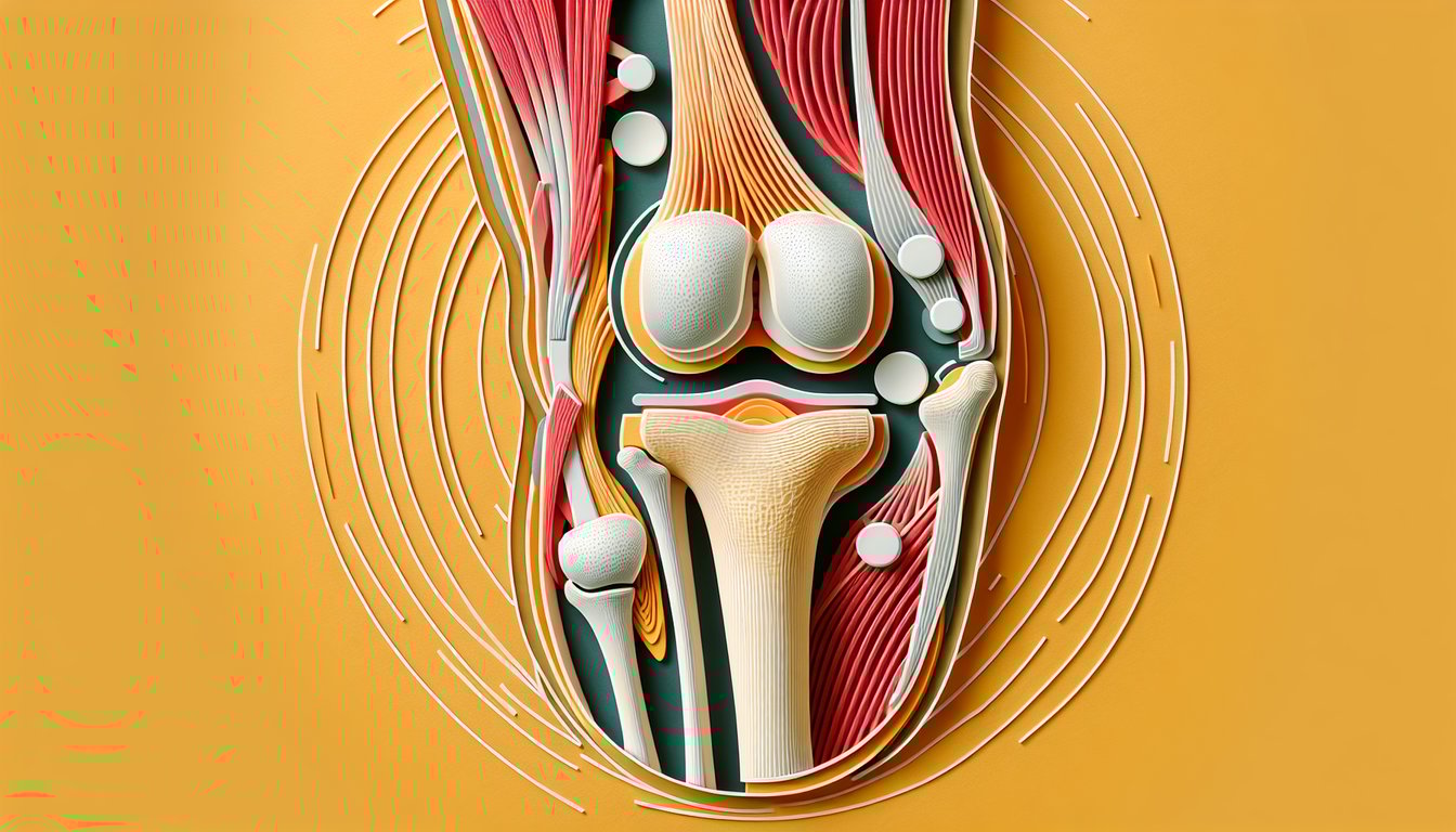
- Bony Structures and Landmarks -
The knee comprises four bones - femur, tibia, patella, and fibula - that form the tibiofemoral and patellofemoral joints. Remember "Find The Perfect Fit" (Femur, Tibia, Patella, Fibula) to nail the knee joint anatomy quiz basics.
- Primary Knee Ligaments -
Four main ligaments - ACL, PCL, MCL, and LCL - stabilize the knee by controlling anterior-posterior and medial-lateral movements. Use the mnemonic "A Pretty Little Love" (Anterior, Posterior, Lateral, Medial) to recall their order and functions.
- Menisci Mechanics -
The medial and lateral menisci are C-shaped fibrocartilage pads that absorb shock and evenly distribute load across the tibiofemoral joint. Note that the lateral meniscus is almost a full circle while the medial is a "C," helping you remember why the medial meniscus tears more often (firmer attachments).
- Joint Classification & Motion -
The knee functions as a hinge joint (tibiofemoral) permitting flexion/extension and as a gliding joint (patellofemoral) for smooth patellar tracking. In your knee ligaments quiz, recall that internal/external rotation occurs only in flexion, critical for the locking mechanism.
- Screw-Home Mechanism -
During the final 20° of extension, the tibia externally rotates ("screws home") to lock the knee, boosting stability with minimal muscular effort. Visualize the tibia "screwing" into extension and "unscrewing" into flexion like a jar lid to master this concept in the ligaments of the knee quiz.