PIC: USLME-DIAGNOSTIC 1

USMLE Diagnostic Quiz
Test your medical knowledge with our comprehensive USMLE diagnostic quiz! This quiz features 50 challenging questions that mimic real exam scenarios and will help you assess your understanding of various medical concepts.
Whether you're preparing for your boards or just want to challenge yourself, this quiz will cover a range of topics, including:
- Dermatology
- Gastroenterology
- Neurology
- Cardiology
- Oncology

A 45-year-old man is brought to the office due a sudden onset of skin lesions and fever. He is unable to eat or drink due to the pain in his mouth and throat. His wife says that he was complaining of a headache, malaise, and joint pain prior to developing the skin lesions. Generally, he has been in good health, other than an episode of sinusitis, for which he was prescribed trimethoprim-sulfamethoxazole 5 days ago. His pulse is 92/min, blood pressure is 110/80 mmHg, respirations are 14/min, and temperature is 36.8°C (98.4°F). On examination, both conjunctivae are inflamed. There is erythema, blistering and ulceration over the oral mucosa. There is an erythematous rash over the trunk and cutaneous lesions over the hands, arms and feet. Some of the lesions are shown in the picture below. What is the most likely diagnosis?
Stevens-Johnson syndrome
Erythema multiforme minor
Staphylococcal scalded skin syndrome
Toxic shock syndrome
Impetigo
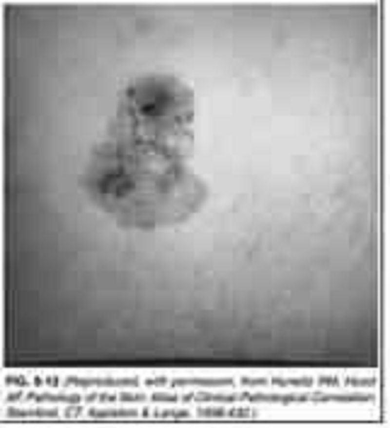
A 45-year-old man presents to the physician’s office for evaluation of a skin lesion on his abdomen. He states that the lesion has been present for 1 year, but has recently enlarged over the last 2 months. The mass is nontender, and he is otherwise asymptomatic. Past history is unremarkable. Examination reveals a 3-cm, pigmented, irregular skin lesion located in the left lower quadrant of the abdomen, as shown in Figure 6-12. Heart, lung, and abdominal examination are normal. There are no palpable cervical, axillary, or inguinal lymph nodes. Chest x-ray and liver fun
Squamous cell carcinoma
Basal cell carcinoma
Merkel cell carcinoma
Melanoma
Keratoacanthoma
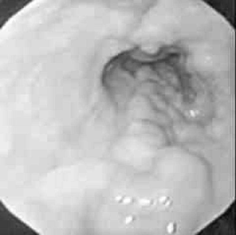
A 45-year-old man with a long history of alcohol intake comes into the emergency room with upper gastrointestinal (UGI) bleeding. Urgent endoscopy reveals the following findings. Which of the following is the most likely diagnosis?
Esophageal varices
Esophageal carcinoma
Foreign body
Tertiary waves
Barrett’s esophagus
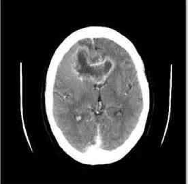
A 45-year-old white male presents with a 4-month history of headaches. The headache is generalized, dull, constant, and worsened by bending, coughing and sneezing. It is unresponsive to simple analgesics, and associated with nausea and vomiting. His wife says he has been acting strangely for the last few months, and she has noted a personality change. The neurological examination is non-focal. Fundoscopy reveals papilledema. His CT scan is shown below. Which of the following is the most likely diagnosis?
Brain abscess
Metastatic brain tumor
Glioblastoma multiforme
Low-grade astrocytoma
Cerebral infarction
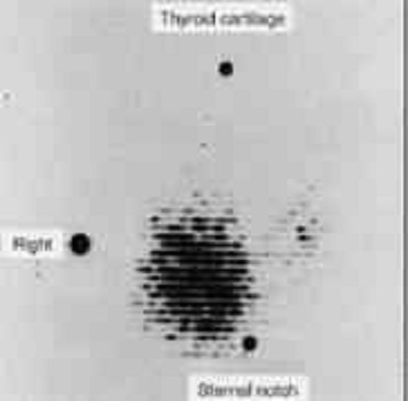
A 45-year-old woman complains to her primary care physician of nervousness, sweating, tremulousness, and weight loss. The thyroid scan shown here exhibits a pattern that is most consistent with which of the following disorders?
Hyper secreting adenoma
Graves’ disease
Lateral aberrant thyroid
Papillary carcinoma of thyroid
Medullary carcinoma of thyroid
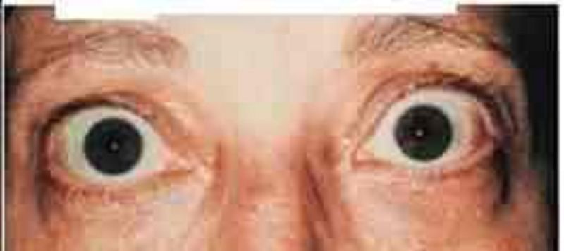
A 46-year-old female complains of a "sandy" sensation in her eyes. Review of systems is notable for a 6 pound weight loss over the last month. A picture of her eyes is shown on the slide below. Which of the following most likely underlies this finding?
High circulating thyroxine level
Periorbital lymphocytic infiltration
Bilateral facial nerve compression
Increased intraocular pressure
Increased intracranial pressure
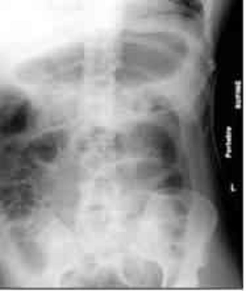
A 46-year-old male is brought to the emergency department after falling on his head and back during a downhill bike race and losing consciousness for 1 minute. He has severe back and abdominal pain. AP and lateral skull films show no abnormalities. Lumbar films show anterior compression wedge fractures of the bodies of L1 and L2. A brace is placed. CT scan of the abdomen shows a mild retroperitoneal bleed and splenic laceration. During the hospitalization he was treated conservatively with analgesics and supportive measures. On hospital day 3, he started to have abdominal distention, pain and nausea. His last bowel movement was 4 days ago and he is not passing gas. His abdomen is distended, tympanic and mildly tender without rebound or guarding. Bowel sounds are absent. An x-ray film of the abdomen is shown below: Which of the following is the most likely diagnosis?
Functional constipation
Paralytic ileus
Large bowel obstruction
Peritonitis
Worsening hematoma
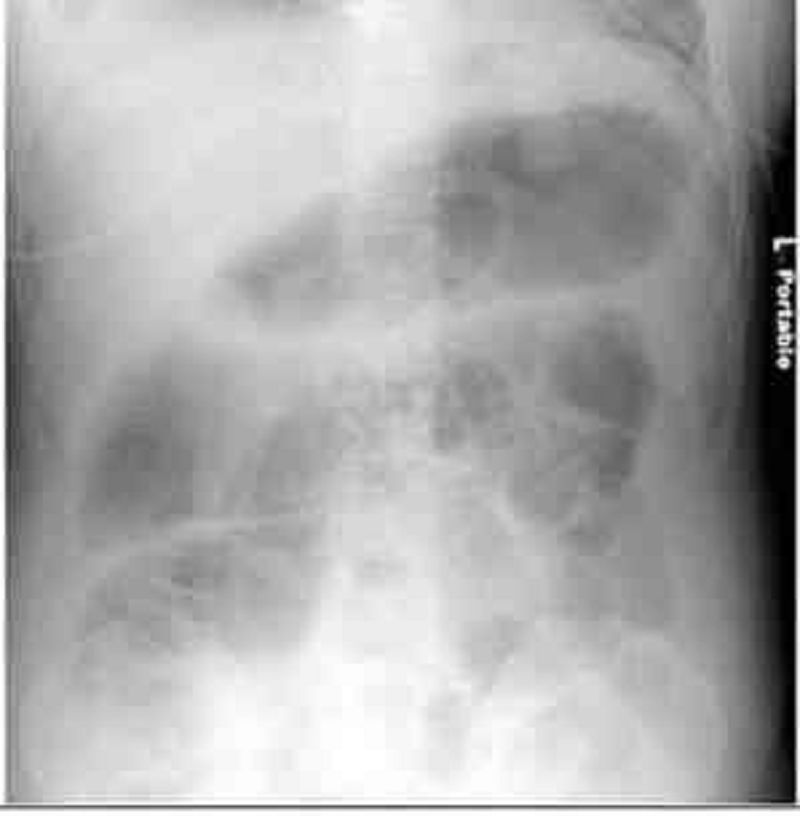
A 46-year-old man is brought to the emergency department after a fall during a downhill bike race. He lost consciousness for approximately 1 minute after the fall. He complains of severe back and abdominal pain. He has no other medical problems. Head computed tomography (CT) scan shows no intracranial bleeding. Lumbar films suggest a compression wedge fracture of the body of L2 vertebra, and a brace is placed. Abdominal CT scan shows a small retroperitoneal bleed and splenic laceration. He is conservatively treated with analgesics and supportive measures. On hospital day three, he complains of abdominal pain and nausea. His abdomen is distended, tympanic, and mildly tender, without rebound or guarding. Bowel sounds are absent. X-ray of the abdomen reveals. Which is the most likely diagnosis?
Erosive gastritis
Expanding retroperitoneal hematoma
Colonic pseudoobstruction
Mesenteric ischemia
Paralytic ileus
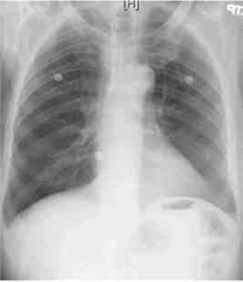
A 46-year-old man presents to the emergency department with difficulty breathing and chest discomfort. His pain worsens with inspiration but does not radiate. He says that he has never had symptoms like this before. His past medical history is unremarkable. He works as a long-haul truck driver. On physical examination, his blood pressure is 110/70 mmHg, his heart rate is 110/min, his respiratory rate is 31/min, and his temperature is 36.7°C (98°F). ECG reveals sinus tachycardia but no ischemic ST-segment or T-wave changes. His chest X-ray is shown below. What is the most likely diagnosis in this patient?
Ascending aortic dissection
Myocardial infarction
Pneumothorax
Pulmonary embolism
Pleural effusion
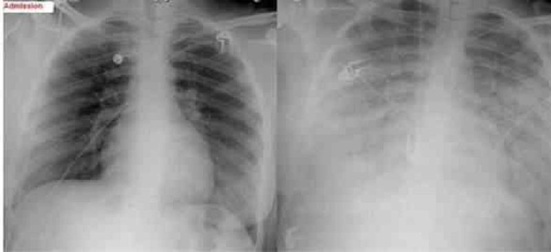
A 47-year-old female, who is a chronic alcoholic, is admitted to the hospital with epigastric pain, nausea, and vomiting. Her serum amylase and lipase levels are significantly elevated and the diagnosis of acute pancreatitis is made. She is maintained nothing by mouth (NPO), and receives intravenous hydration and narcotic analgesics. On the second day of hospitalization she develops progressive shortness of breath. Her temperature is 37.2°C (98.9°F), blood pressure is 110/66 mm Hg, pulse is 110/min, and respirations are 24/min. Her oxygenation is measured at 84% on 100% non-rebreather mask and the decision is made to intubate. Since the time of admission, she has received 5 liters of normal saline and has produced 3 liters of urine output. On examination, there is no evidence of jugular venous distention. Chest auscultation reveals diffuse bilateral crackles. Auscultation of the heart reveals normal heart sounds with no murmurs. A chest x-ray from the time of admission and one from the time of intubation are shown below. Based on these findings, what is the most likely diagnosis in this patient?
Acute respiratory distress syndrome
Hospital acquired pneumonia
Iatrogenic volume overload
Congestive heart failure from myocardial infarction
Alcoholic cardiomyopathy
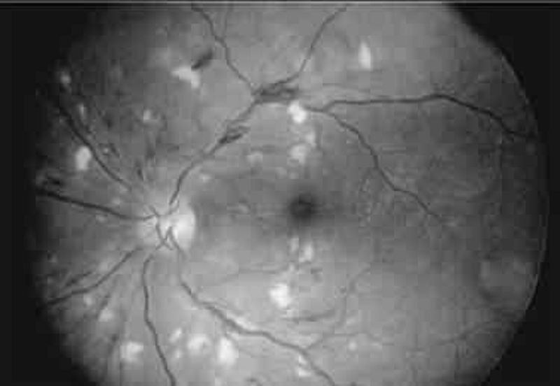
A 47-year-old woman develops accelerated hypertension (blood pressure 210/105 mmHg) but no clinical symptoms except frequent headaches. Which of the following findings are most likely on examination of the fundii?
Retinitis obliterans
Cotton wool spots
Retinal detachment
Optic atrophy
Foveal blindness
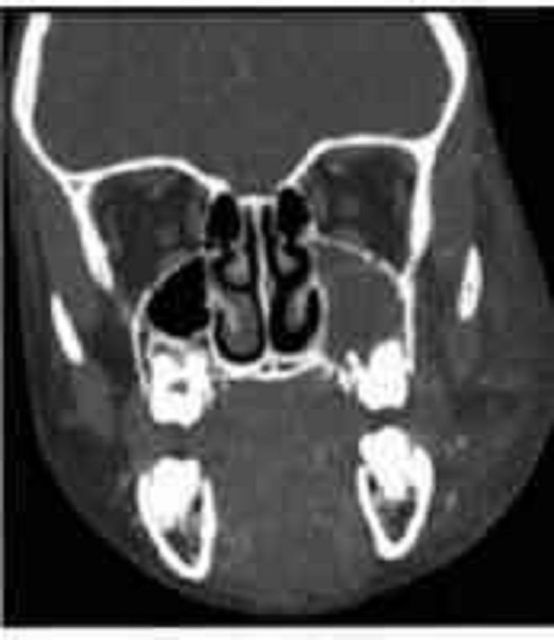
A 5-year-old girl is brought to the physician with low grade fever and rhinorrhea. Her symptoms began ten days ago. She has also had persistent purulent rhinorrhea, nasal congestion, and a dry cough during the day that worsens at night. Her symptoms do not seem to be improving. On examination, the child has erythema and swelling of the nasal turbinates with purulent nasal drainage. She has evidence of drainage in the posterior pharynx as well. The remainder of her examination is unremarkable. Computed topography of her face is shown below. Which of the following is the most common predisposing factor for her condition?
Allergic rhinitis
Septal deformities
Adenoidal hypertrophy
Environmental mucosal irritants
Viral upper respiratory infection
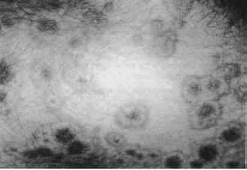
A 50-year-old woman develops pink macules and papules on her hands and forearms in association with a sore throat. The lesions are target like, with the centers a dusky violet. What causes of this disorder are most likely in this patient?
Tampons and superficial skin infections
Drugs and herpesvirus infections
Rickettsial and fungal infections
Anxiety and emotional stress
Harsh soaps and drying agents
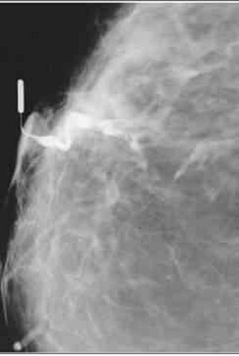
A 51-year-old woman presents to the physician’s office with a 2-month history of a right breast blood tinged nipple discharge. Past history is unremarkable. Family history is positive for postmenopausal breast cancer in a maternal grandmother. Examination reveals no palpable masses or regional adenopathy, but a serous discharge is easily elicited from a single duct in the right breast. Bilateral mammograms show no abnormalities. Cytology from the discharge was not diagnostic. A ductogram was ordered, and the results are shown: Which of the following is the most likely diagnosis?
Invasive carcinoma
Intraductal carcinoma
Intraductal papilloma
Fibrocystic disease
duct ectasia
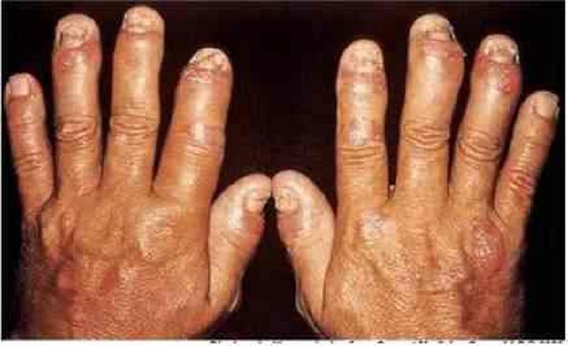
A 52-year-old male presents with a long history of joint pain. He describes pain and stiffness of the small joints of his hand that is worse in the morning and can last several hours. He also complains of occasional digit swelling. A picture of the patient's hands is shown on the slide below. Which of the following is the most likely diagnosis?
Enteropathic arthritis
Rheumatoid arthritis
Psoriatic arthritis
Crystalline arthritis
Sarcoidosis
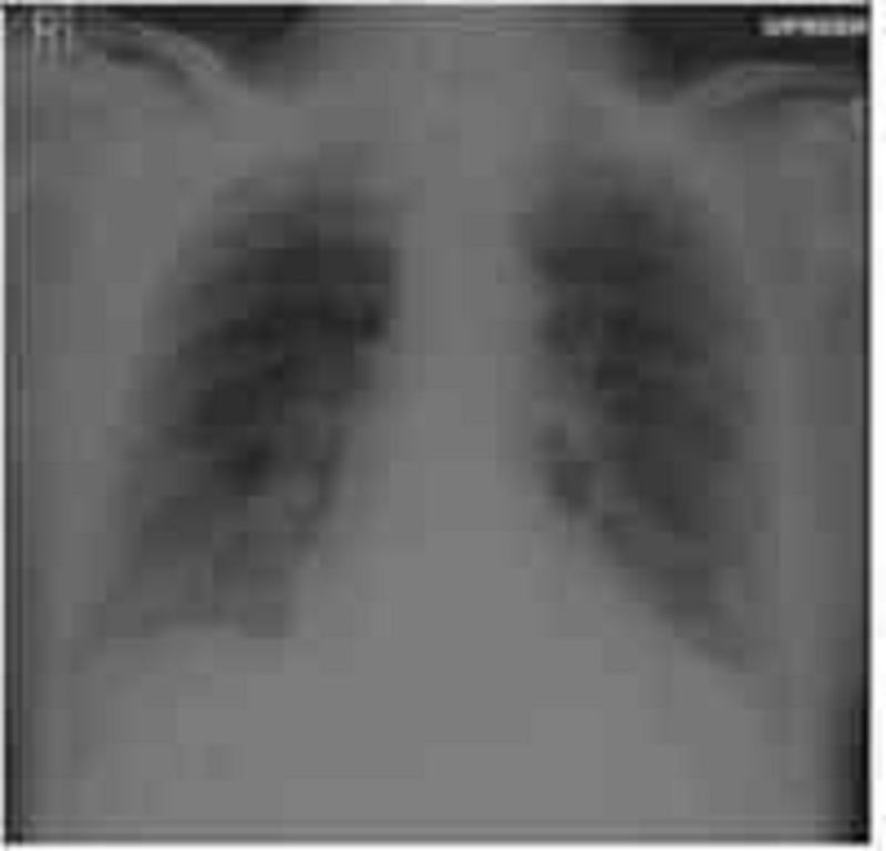
A 53-year-old male comes to the emergency department complaining of sudden onset intense, stabbing epigastric pain. He also vomited once and a dull, aching pain then spread through his entire abdomen. He has had nonspecific epigastric pain for several months and saw a physician one month ago. He also has a history of constipation, type II diabetes mellitus and hyperlipidemia. He has smoked one and a half packs of cigarettes daily for 26 years. He drinks 4 oz of alcohol daily. His temperature is 38.3C (100.4F), blood pressure is 160/95 mm Hg, pulse is 100/min and respirations are 26/min. The entire abdomen is tender to palpation with rebound, but there is no guarding. No masses are palpable, and Murphy's sign elicits mild pain. Rectal examination shows no abnormalities. Abdominal ultrasound performed 2 weeks ago showed stones in the gall bladder. Upright chest x-ray is shown below: Which of the following is the most likely diagnosis in this patient?
Acute cholecystitis
Acute alcoholic pancreatitis
Acute gallstone pancreatitis
Perforated peptic ulcer
Perforated diverticulitis
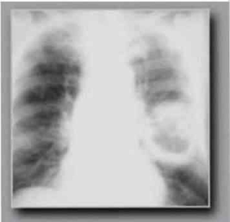
A 54-year-old black male from the southeast USA presents to you with complaints of generalized malaise, fever, and a cough. He claims that he has had intermittent hemoptysis for the past six months. He denies smoking and has never had tuberculosis. Examination is unremarkable and his chest x-ray is shown below. On changing position, you notice that the part of the lesion seen on x-ray also moves. The most likely diagnosis is?
Lung abscess
Pulmonary embolism
Aspergilloma
Histoplasmosis
Bronchiectasis
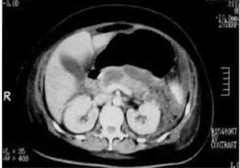
A 54-year-old male presents to the emergency department with a 1-week history of abdominal pain. His other symptoms are nausea, vomiting, low-grade fever, and loss of appetite. He does not use alcohol. He has a seizure disorder, for which he takes a "prescription drug." X-ray films of his chest and abdomen show no abnormalities. His abdominal CT scan is shown below. Which of the following is the most likely explanation for this patient's abdominal symptoms?
Gall bladder pathology
Kidney pathology
Pancreas pathology
Air in the stomach
Liver pathology
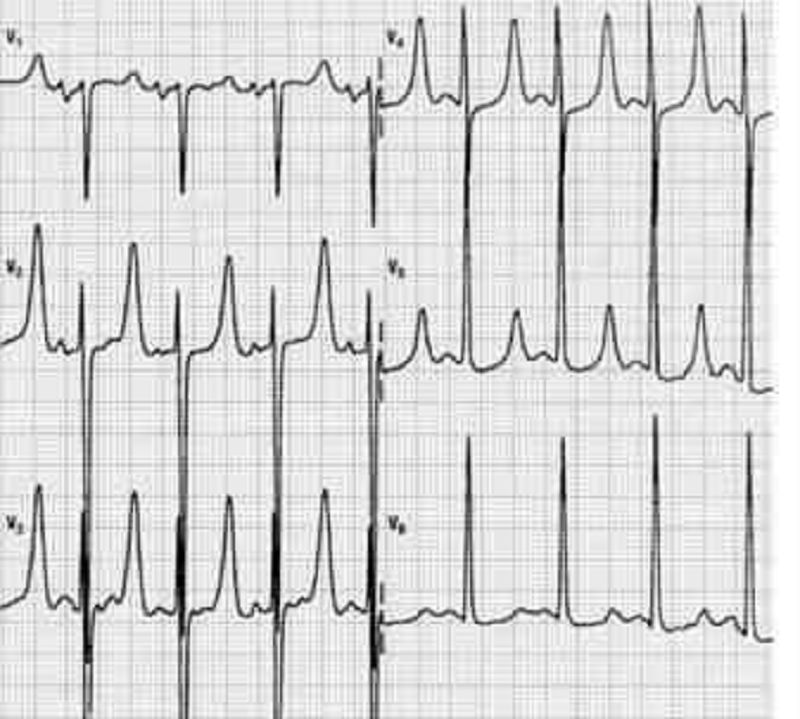
A 54-year-old woman presents to the ED because of a change in behavior at home. For the past 3 years, she has end-stage renal disease requiring dialysis. Her daughter states that the patient has been increasingly tired and occasionally confused for the past 3 days and has not been eating her usual diet. On examination, the patient is alert and oriented to person only. The remainder of her examination is normal. An initial 12-lead ECG is performed as seen on the following page. Which of the following electrolyte abnormalities best explains these findings?
Hypokalemia
Hyperkalemia
Hypocalcemia
Hypercalcemi
Hyponatremia
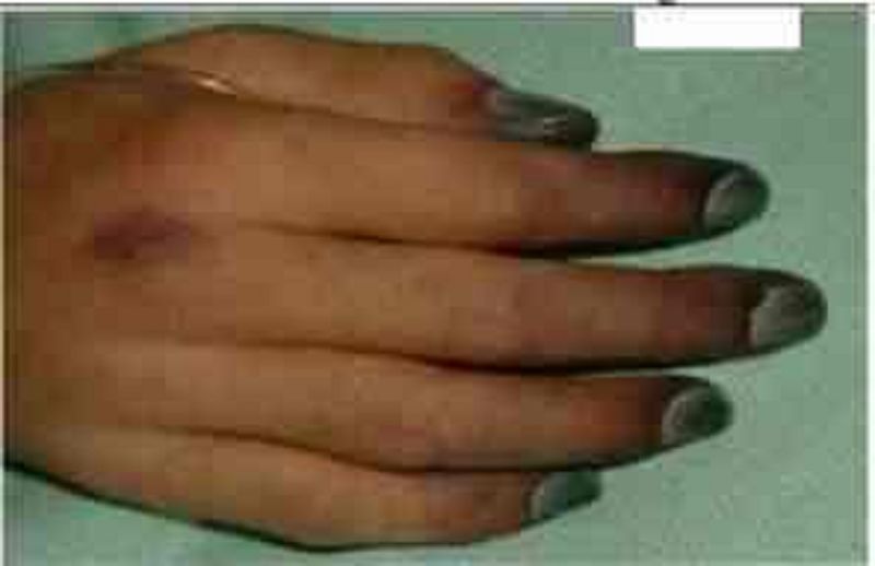
A 55-year-old male is admitted to the ICU after being involved in a motor vehicle accident. He requires exploratory laparotomy for suspected bowel perforation. Two days after the surgery he remains hypotensive and requires both aggressive intravenous fluids and vasopressors to maintain his blood pressure. On physical examination, you note the fingertip changes pictured below. All four extremities feel cold to touch. Which of the following is most likely responsible?
Septic emboli
Raynaud's phenomenon
Norepinephrine-induced vasospasm
Cholesterol emboli
Superior vena cava syndrome
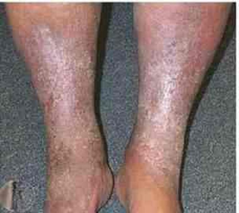
A 55-year-old man comes to the physician because of chronic leg problems. He has had multiple medical problems and is unable to get good medical care due to lack of insurance. A photograph of his legs is shown below. Which of the following is the most likely cause of his condition?
Arterial thrombosis
Arterial spasm
Venous hypertension
Peripheral neuropathy
Posterior spinal cord lesion
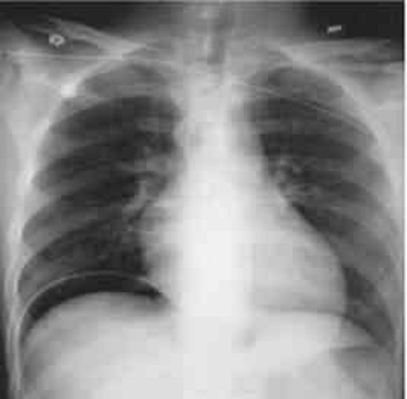
A 55-year-old man presents to the ED complaining of mild diffuse abdominal pain. He states that he underwent a routine colonoscopy yesterday and was told “everything is fine.” The pain began upon waking up and is associated with some nausea. He denies fever, vomiting, diarrhea, and rectal bleeding. His BP is 143/71 mm Hg, HR is 87 beats per minute, temperature is 98.9°F, and RR is 16 breaths per minute. His abdomen is tense but only mildly tender. You order baseline laboratory tests. His chest radiograph is seen below. Which of the following is the most likely diagnosis?
Ascending cholangitis
Acute pulmonary edema
Acute liver failure
Pancreatitis
Pneumoperitoneum
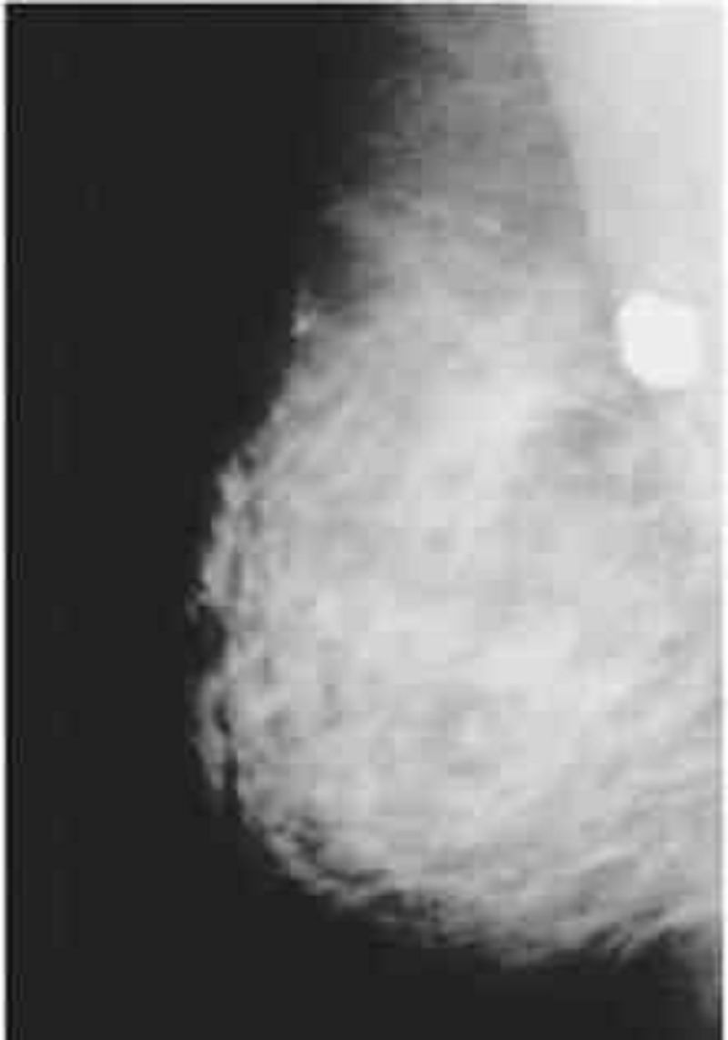
A 55-year-old-woman presents to the physician’s office for evaluation of mammographic findings on a screening mammogram. She denies any breast masses, nipple discharge, pain, or skin changes. Past history is pertinent for insulin-dependent diabetes. Family history is positive for postmenopausal breast cancer in her mother. She has a normal breast examination and no axillary adenopathy. A mediolateral oblique (MLO) view of the right breast is shown: Which of the following is the most likely diagnosis?
milk of calcium
Lobular carcinoma in situ (LCIS) with or without an invasive component
ductal carcinoma in situ (DCIS) with or without an invasive component
Involuting fibroadenoma
phyllodes tumor

A 56-year-old diabetic female comes to the clinic with complaints of dizziness which has been going on for 3 weeks. She denies any dyspnea or diaphoresis. She says her blood glucose is well controlled and denies any allergy. Her BP is 155/90 mmHg. Her chest-x ray is unremarkable and her blood work is normal. The ECG is recorded below. What is the most likely diagnosis?
Mobitz type I heart block
Mobitz type II heart block
Complete heart block
Atrial fibrillation
First degree heart block
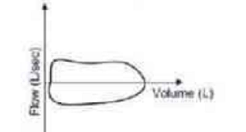
A 56-year-old woman is brought to the hospital from a local restaurant after suddenly becoming short of breath. Her flow-volume loop is shown below. Which of the following is the most likely cause of her symptoms?
Asthma attack
Pneumothorax
Pulmonary edema
Laryngeal edema
Panic attack
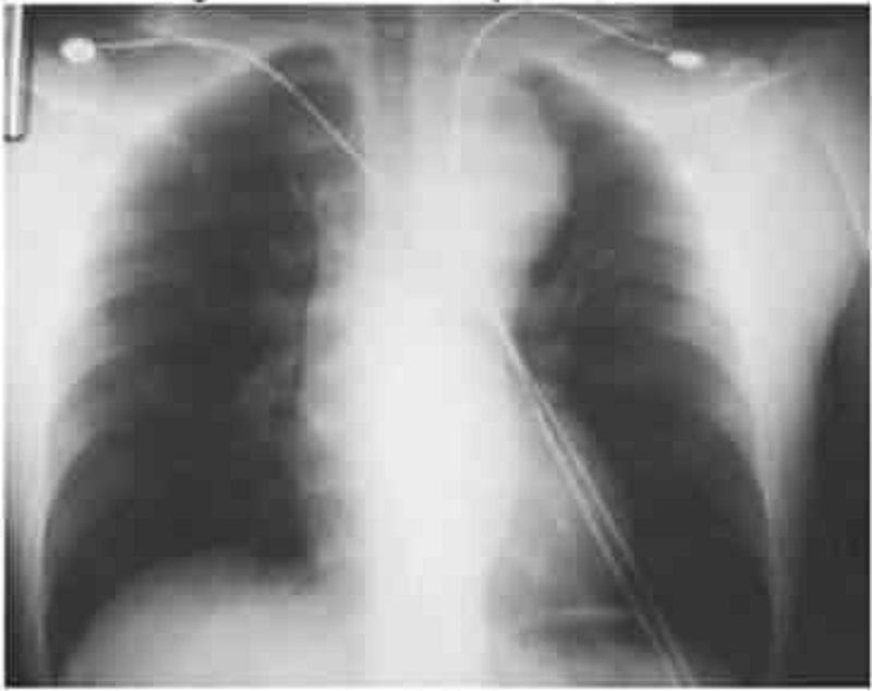
A 58-year-old man with hypertension is brought to the emergency room after sudden onset chest pain that radiates to his back and arms. He is in moderate distress with a blood pressure of 160/90 mmHg in the left arm and 120/70 mm Hg in the right arm. Cardiac examination reveals a soft second heart sound and a murmur of aortic insufficiency. His ECG shows sinus tachycardia but no acute ischemic changes, and the chest x-ray (CXR). Which of the following is the most appropriate next step in confirming the diagnosis?
Coronary angiography
Transthoracic echocardiography
Computerized tomography (CT) chest
Exercise stress test
Cardiac troponin level
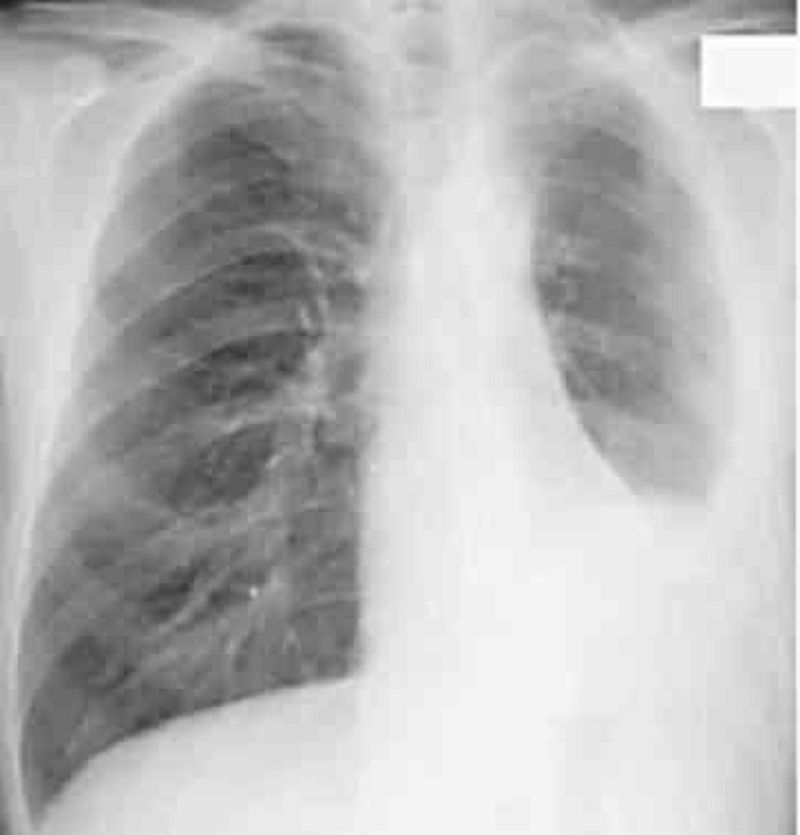
A 58-year-old steam pipe worker presents with a vague ache in the left chest and mild dyspnea of several months’ duration. There is dullness on percussion of the left chest associated with diminished breath sounds. His CXR is shown in Fig. Which of the following is the most likely diagnosis?
Pleural metastases
Paget’s disease
Mesothelioma and asbestosis
Pleural effusion
Multiple myelom
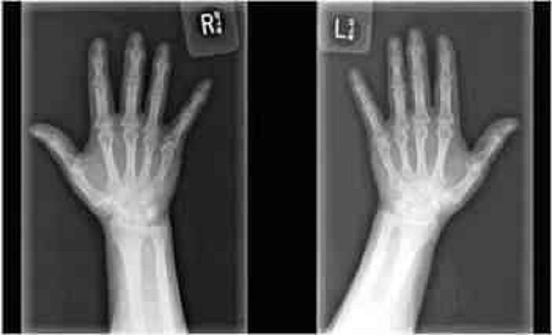
A 60-year-old Caucasian woman comes to the physician because of joint pains in both hands. Her other medical problems include obesity and gastroesophageal reflux disease. She does not use tobacco, alcohol, or drugs. Family history is not significant. Her medications include omeprazole and acetaminophen. Her vital signs are within limits. X-ray of the joints is shown below. Which of the following is the most likely diagnosis?
Rheumatoid arthritis
Systemic lupus erythematosus
Osteoarthritis
Reactive arthritis
Gouty arthritis
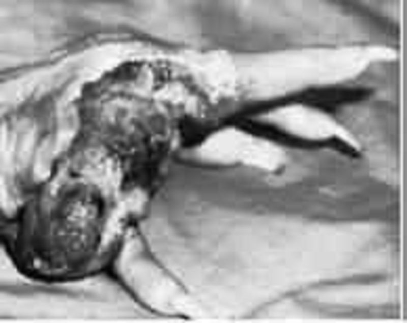
A 60-year-old woman presents with the skin lesion shown here. She reports a history of a burn injury to the hand while cooking a few years ago. She reports the wound has never healed completely. You are concerned about the skin lesion and perform a punch biopsy. Which of the following is the most accurate diagnosis given the patient’s history?
Basal cell carcinoma
Malignant melanoma
Erythroplasia of Queyrat
Bowen disease
Marjolin ulcer
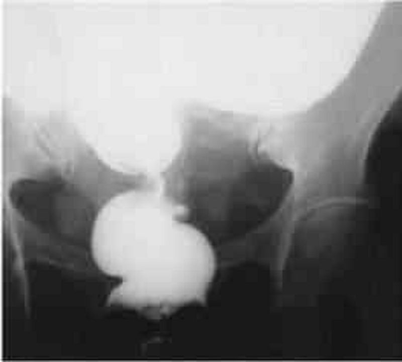
A 62-year-old woman presents to the physician’s office with complaints of constipation. She has had constipation for the last 6 months, which has worsened over the last month, associated with mild bloating. She noted that her stool has become “pencil thin” in the last month, with occasional blood, but she continues to have bowel movements daily. Past history is unremarkable. Examination reveals normal vital signs and heart and lung examination. Abdominal examination reveals mild fullness, especially in the lower quadrants. Rectal examination shows no rectal masses, but the stool is hematest positive. A barium xray is obtained, and one view is shown in Figure 6-11. Which of the following is the most likely diagnosis?
Crohn’s disease
Ischemia with stricture
rectal carcinoma
Sigmoid volvulus
Diverticulitis with colovesical fistula

A 63-year-old female presents to your clinic complaining of palpitations. For the past 3 weeks, she has noticed pounding of her heart that comes and goes. Her symptoms are more frequent at night. Her only medicine is insulin for diabetes mellitus. On physical examination, she is alert and oriented, and in no distress. Her EKG is shown below. Which of the following best accounts for this patient's symptoms?
Sinus arrhythmia
Irregularly irregular atrial activation
Variable AV node conduction
Atrial ectopy
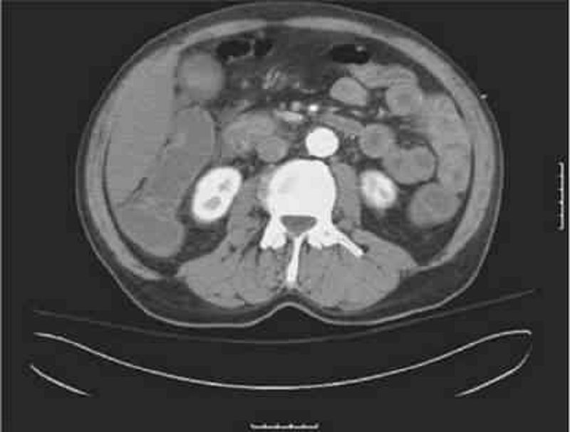
A 63-year-old man is brought to the ED by EMS complaining of severe abdominal pain that began suddenly 6 hours ago. His BP is 145/75 mm Hg and HR is 105 beats per minute and irregular. On examination, you note mild abdominal distention and diffuse abdominal tenderness without guarding. Stool is heme positive. Laboratory results reveal WBC 12,500/μL, haematocrit 48%, and lactate 4.2 U/L. ECG shows atrial fibrillation at a rate of 110. A CT scan is shown below. Which of the following is the most likely diagnosis?
Abdominal aortic aneurysm
Mesenteric ischemia
Diverticulitis
SBO
Crohn disease
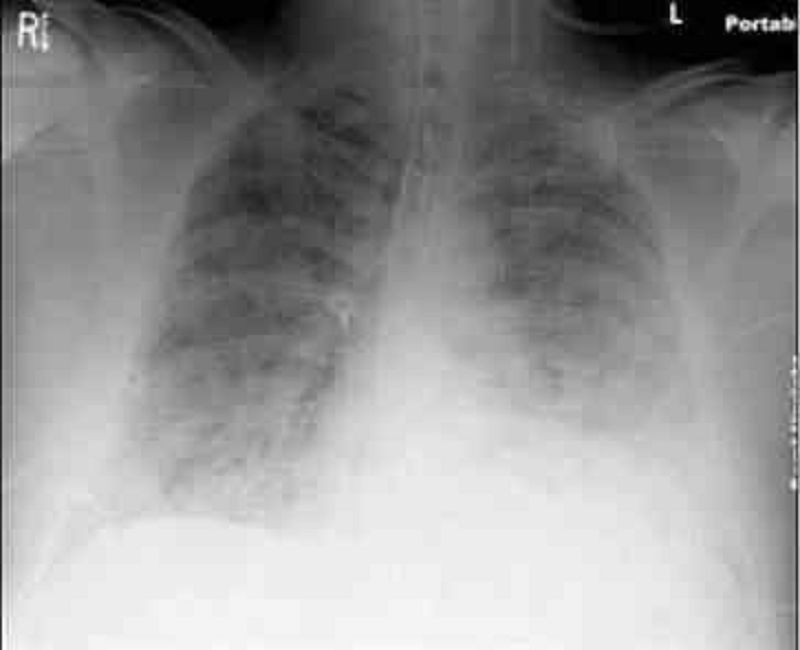
A 64-year-old male is admitted to the hospital with abdominal pain, abdominal distention, and confusion. Upon arrival his blood pressure is 90/60 mmHg and pulse is 120/min. On physical examination, his abdomen is tender, distended, and rigid with positive rebound tenderness. His past medical history is significant for rheumatic fever as a child, hypertension, coronary artery disease and atrial fibrillation. He receives a total of 6 liters of normal saline and undergoes emergent laparotomy. Postoperatively he complains of shortness of breath. His respiratory rate is 34/min. He is emergently intubated because of poor oxygenation. His chest x-ray is shown below. This film is compared to a chest x-ray performed one week earlier, which was within normal limits. Currently, the pulmonary capillary wedge pressure is 8 mmHg. Which of the following is the most likely cause of his current condition?
Idiopathic pulmonary fibrosis
Mitral stenosis
Acute respiratory distress syndrome
Left ventricular systolic dysfunction
Iatrogenic fluid overload
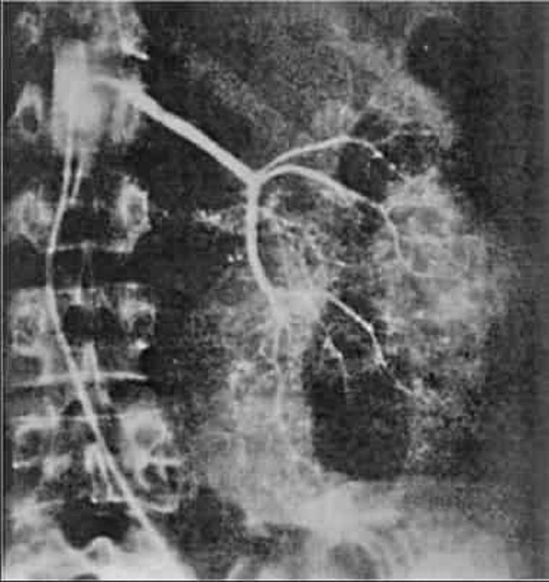
A 64-year-old man is admitted for hematuria after slipping on an icy pavement. His physical examination is normal. A selective angiogram of the left kidney is shown in Fig. Which of the following is the most likely diagnosis?
Renal cell carcinoma
Kidney contusion and laceration
Transitional cell carcinoma
Renal hamartoma
Renal hemangioma
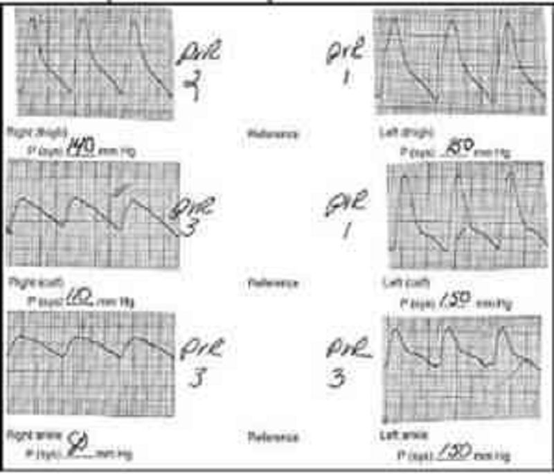
A 65-year-old male cigarette smoker reports onset of claudication of his right lower extremity approximately 3 weeks previously. He can walk 3 blocks before the onset of claudication. Physical examination reveals palpable pulses in the entire left lower extremity, but no pulses are palpable below the right groin level. Non-invasive flow studies are obtained and are pictured here. What is the level of the occlusive process in this patient?
Right anterior tibial artery
Right superficial femoral artery
Right profunda femoris artery
Right external iliac artery
Right internal iliac artery
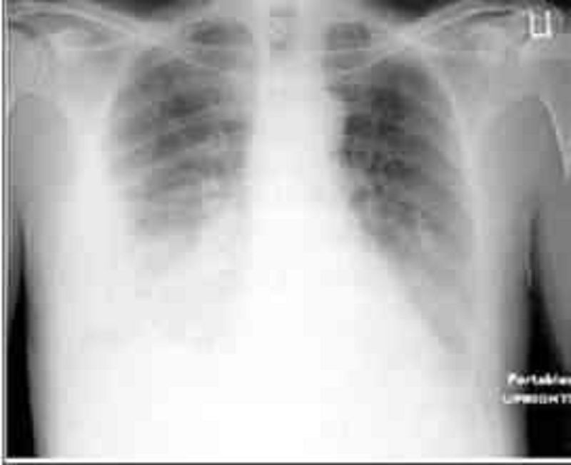
A 65-year-old male comes to the emergency department with severe shortness of breath. The symptoms started one week ago with fever and a non-productive cough. His past medical history is significant for coronary artery disease with bypass surgery two years ago, hypertension and diabetes mellitus. His temperature is 38.9°C (102°F), blood pressure is 160/70 mm Hg, pulse is 110/min, and respirations are 26/min. Physical examination reveals decreased breath sounds over the right lower lung base. His chest X-ray is shown on the slide below. Which of the following is the most likely cause of this patient's current complaints?
Bronchopleural fistula
Lung abscess
Empyema
Pneumothorax
Pulmonary infarction
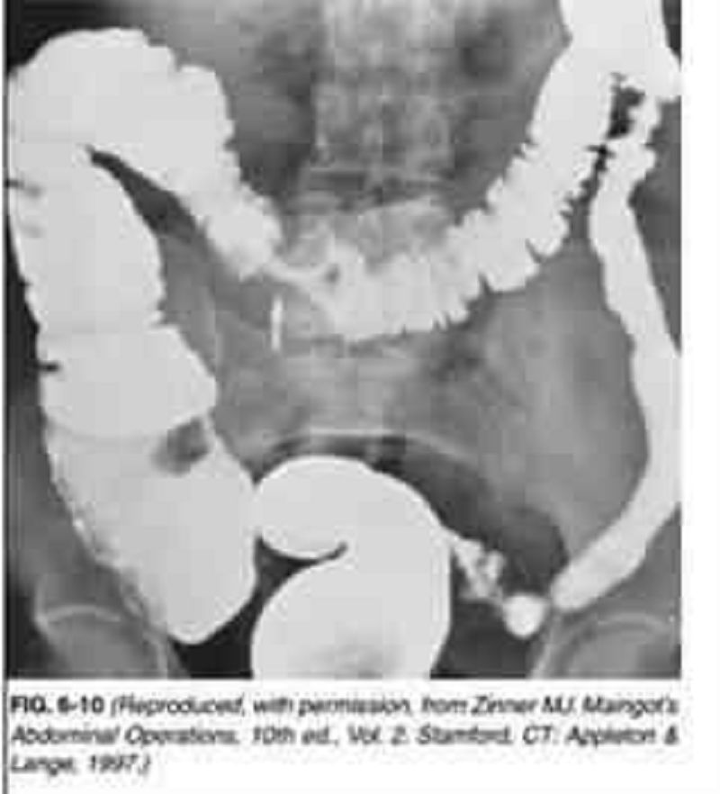
A 65-year-old man presents to the physician’s office for his yearly physical examination. His only complaint relates to early fatigue while playing golf. Past history is pertinent for mild hypertension. Examination is unremarkable except for trace hematest-positive stool. Blood tests are normal except for a hematocrit of 32. A UGI series is performed and is normal. A barium enema is performed, and one view is shown in Figure 6-10. Which of the following is the most likely diagnosis?
Diverticular disease
Colon cancer
Lymphoma
Ischemia with stricture
Crohn’s colitis with stricture
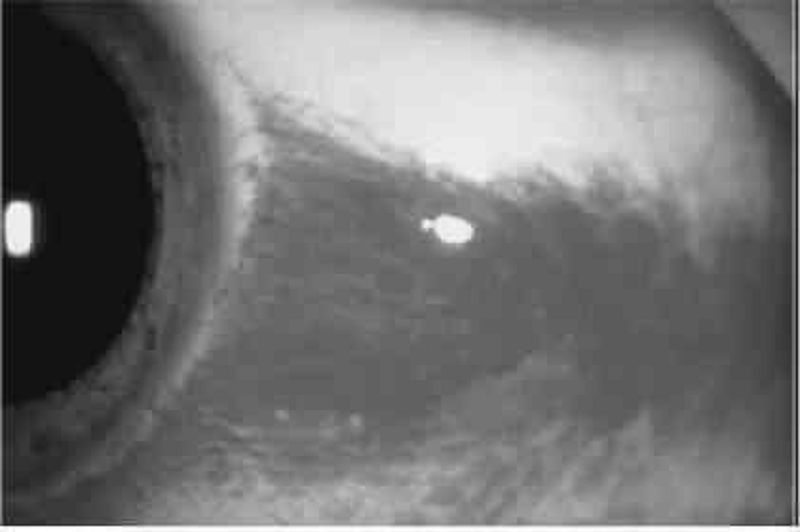
A 65-year-old man with a history of diabetes, hypertension, coronary artery disease, and atrial fibrillation presents with loss of vision in his left eye since he awoke 6 hours ago. The patient denies fever, eye pain, or eye discharge. On physical examination of the left eye, vision is limited to counting fingers. His pupil is 3 mm and reactive. Extraocular movements are intact. Slit-lamp examination is also normal. The dilated funduscopic examination is shown below. Which of the following is the most likely diagnosis?
Retinal detachment
Central retinal artery occlusion
Central retinal vein occlusion
Vitreous hemorrhage
Acute angle-closure glaucoma
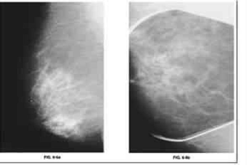
A 65-year-old woman presents to the physician’s office for evaluation of an abnormal screening mammogram. She denies any breast masses, nipple discharge, pain, or skin changes. Past history is pertinent for hypertension. Family history is positive for postmenopausal breast cancer in a sister. She has a normal breast examination and no axillary adenopathy. The remainder of her examination is unremarkable. An MLO view of the right breast is shown in Figure 6-6a along with a magnification view of the craniocaudal (CC) film (Figure 6-6b). Which of the following is the most likely diagnosis?
Milk of calcium
LCIS with or without an invasive component
DCIS with or without an invasive component
involuting fibroadenoma
Phyllodes tumor
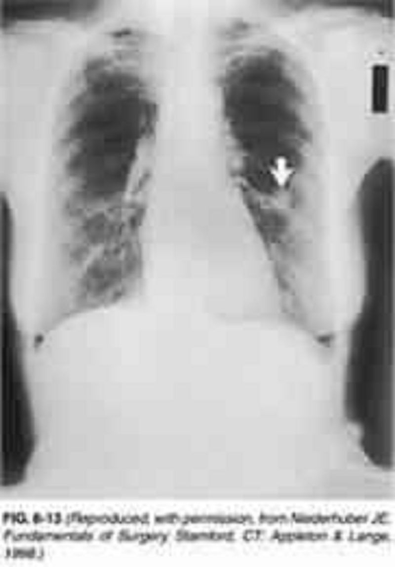
A 65-year-old woman presents to the physician’s office for her yearly physical examination. She has no complaints except for a recent 10-lb weight loss. Past history is pertinent for a 40 pack-year smoking history, hypertension, asthma, and hypothyroidism. Examination reveals a thin woman with normal vital signs and unremarkable heart and abdominal examinations. Lung examination reveals mild wheezing and a few bibasilar rales. A chest x-ray is obtained and is shown in Figure 6-13. A chest x-ray obtained 3 years ago was normal. Yearly laboratory tests including a CBC, electrolytes, and lipid panels are normal. Which of the following is the most likely diagnosis?
Small cell lung cancer
tuberculosis
nonsmall cell lung cancer
Hamartoma
Abscess

A 65-year-old woman presents to the physician’s office with a 6-month history of epigastric discomfort, poor appetite, and 10-lb weight loss. Past history is pertinent for hypertension, diabetes, a 30 pack-year smoking history, and occasional alcohol intake. Examination is unremarkable except for mild epigastric tenderness to deep palpation. An abdominal ultrasound reveals cholelithiasis, and one view of a UGI x-ray series is shown in Figure 6-8. Which of the following is the most likely diagnosis?
cholecystoenteric fistula
Duodenal ulcer
Gastric ulcer
Gastric diverticulum
Duodenal diverticulum
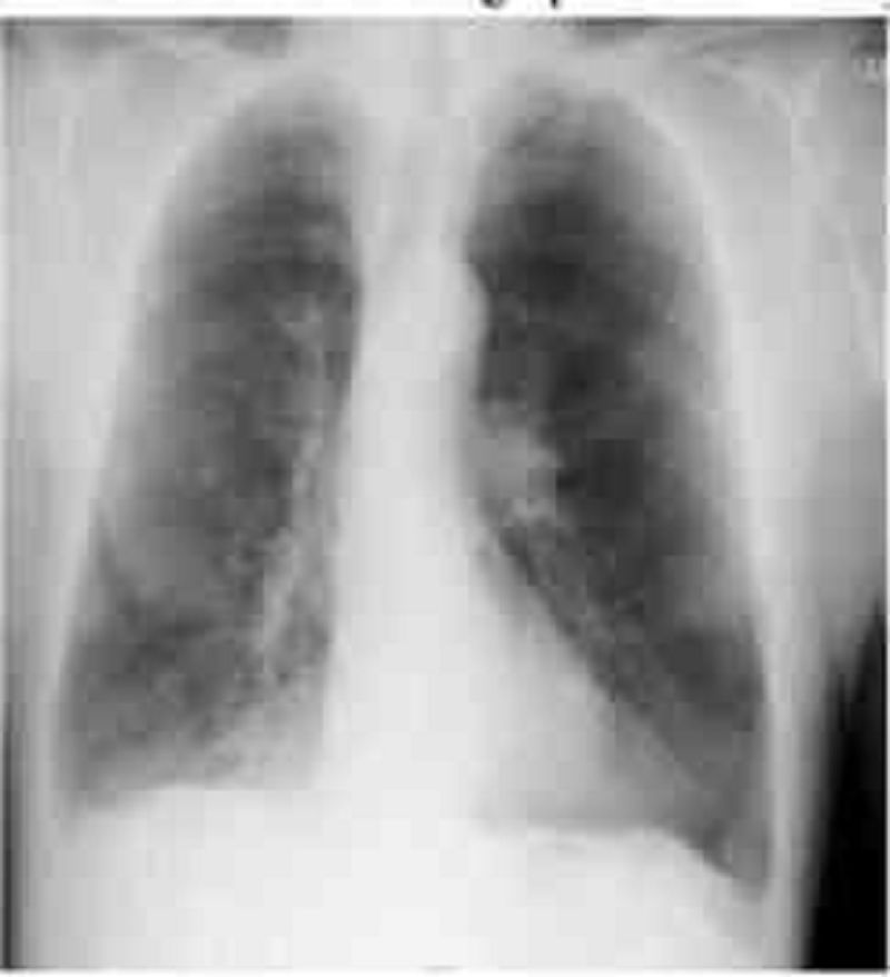
A 69-year -old Caucasian man presents with a two-day history of increasing shortness of breath and lower extremity edema. He is currently short of breath at rest and has an occasional cough. There is no past history of hypertension or ischemic heart disease. He reports drinking half a bottle of vodka daily and has smoked 1 pack of cigarettes per day for 45 years. His blood pressure is 160/90 mm Hg, pulse is 90/min, and oxygen saturation is 90% on room air. JVP is elevated and auscultation of his heart reveals faint heart sounds. The liver span is 18 cm and ascites is also present. No rales are heard in the lungs. There is 3+ lower extremity pitting edema up to the knees. The chest radiograph is shown below. Which of the following is the most likely diagnosis?
Alcoholic cirrhosis
Coronary artery disease
Cardiac tamponade
Metastatic carcinoma of the liver
Cor pulmonale
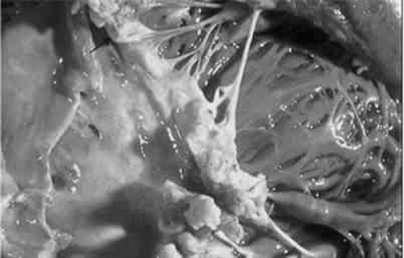
A 69-year-old man with rheumatic heart disease presents to the emergency department complaining of a fever and weakness on his left side. On physical examination the patient is weak in his left upper extremity and he draws only the right half of a clock. Shortly after his presentation, the patient dies, and an autopsy is performed. A gross view of the patient’s heart is shown in the image. Which of the following is a risk factor for the type of lesion pictured?
Coronary artery disease
Hypertension
Mitral valve prolapse
Prolonged bedrest
Prosthetic valve replacement
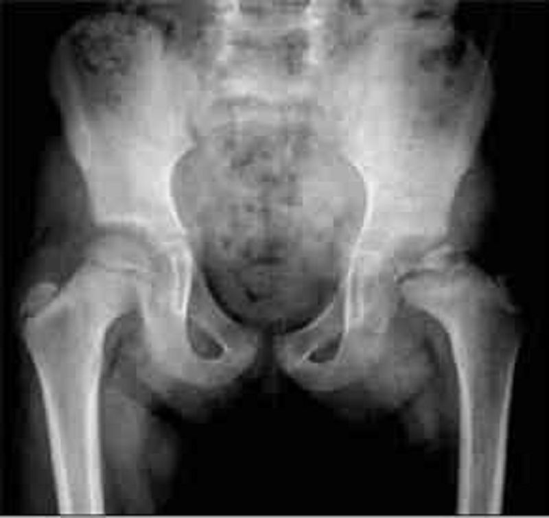
A 7-year-old boy has been complaining of left hip pain for the past 8 months. Over recent weeks, he has developed a limp. When you examine his gait, you note that he takes short steps with his left leg. On physical examination, his left hip has significantly limited range of motion, and there is atrophy of the left proximal thigh muscle. X-ray of the patient's pelvis is shown below: W hich of the following is most likely responsible for this patient's condition?
Slipped epiphysis
Bone infection
Osteonecrosis
Muscle dystrophy
Synovitis

A 7-year-old male is brought to the emergency department for a suspected femur fracture. He has had multiple fractures in the past after minor trauma. Today, his mother states that he was running and fell. He complained of pain in his thigh after he fell. His examination is remarkable for tenderness to palpation and slight deformity of his right proximal thigh. He has decreased muscle tone throughout. His eye examination is shown below. Which of the following is the most likely associated finding?
Aortic root dilatation
Horseshoe kidney
Opalescent teeth
Mental retardation
Ash leaf macules

A 70-year-old Caucasian male presents to your office for evaluation of skin lesions on his forehead. On physical exam you find that these papules have a sandpaper texture by palpation. The lesions are illustrated in the slide below. Which of the following is the most likely diagnosis in this patient?
Psoriasis
Seborrheic keratosis
Actinic keratosis
Atopic dermatitis
Pityriasis rosea
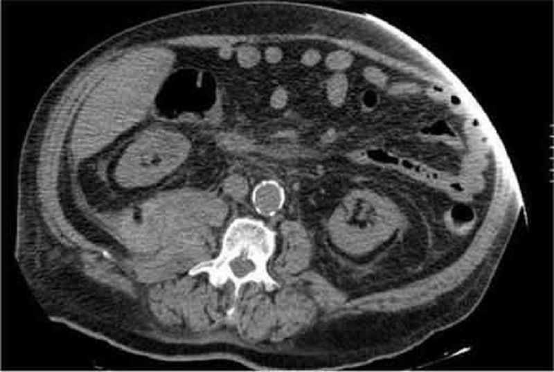
A 70-year-old male presents to the emergency room complaining of weakness, dizziness and back pain. He denies nausea, vomiting, diarrhea, chest pain, palpitations, shortness of breath, urinary symptoms, or black stools. His past medical history is significant for diabetes mellitus, diabetic nephropathy and retinopathy, hypertension, atrial fibrillation and chronic leg cellulitis. He takes warfarin for chronic anticoagulation. On physical examination, his blood pressure is 139/75 mmHg and his heart rate is 110 and irregular. His WBC count is 10,500/mm3, hemoglobin level is 7.0 mg/dl and platelet count is 170,000/mm3. An abdominal CT image is shown on the slide below. Which of the following is the most likely diagnosis?
Renal cell carcinoma
Vertebral fracture
Retroperitoneal hematoma
Hydronephrosi
Mesenteric ischemia
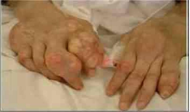
A 71-year-old female is brought to your clinic by her daughter with a complaint of severe pain in her fingers. Her daughter says, "Mom has horrible problems with her joints and she has never tried to get help". The patient adds that her fingers have been swollen and painful for a few weeks. She claims that she had a similar condition in her foot last year. She was given a pain pill, but it was ineffective. She takes a water pill for her blood pressure. What is the most likely diagnosis in this patient?
Rheumatoid nodules
Gouty arthritis
Severe osteoarthritis
Bone tumor
Severe psoriatic arthritis

A 73-year-old man is seen in the ED for abdominal pain, nausea, and vomiting. His symptoms have progressively worsened over the past 2 to 3 days. The pain is diffuse and comes in waves. He denies fever or chills, but has a history of constipation. He reports no flatus for 24 hours. Physical examination is notable for diffuse tenderness and voluntary guarding. There is no rebound tenderness. An abdominal radiograph is seen below. Which of the following is the most likely diagnosis?
Constipation
SBO
Cholelithiasis
Large bowel obstruction
Inflammatory bowel disease
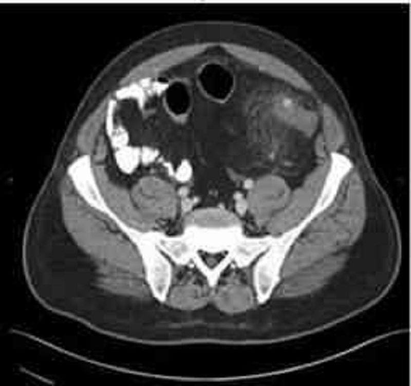
A 74-year-old man presents to the emergency department with abdominal pain. The pain is deep and aching and is localized to the left lower quadrant. The man reports multiple episodes of diarrhea over the preceding week. He also reports having multiple similar episodes of abdominal pain in the past. On physical examination he is febrile and has tenderness to palpation of the left lower quadrant. His WBC count is 23,000/mm³. Results of CT are shown in the image. Which of the following is the most likely diagnosis?
Angiodysplasia
Carcinoid syndrome
Carcinoma of the colon
Diverticulitis
Infectious colitis
{"name":"PIC: USLME-DIAGNOSTIC 1", "url":"https://www.quiz-maker.com/QPREVIEW","txt":"Test your medical knowledge with our comprehensive USMLE diagnostic quiz! This quiz features 50 challenging questions that mimic real exam scenarios and will help you assess your understanding of various medical concepts.Whether you're preparing for your boards or just want to challenge yourself, this quiz will cover a range of topics, including:DermatologyGastroenterologyNeurologyCardiologyOncology","img":"https:/images/course7.png"}
More Quizzes
USMLE_Basic V
1507578
QCU/DES/USMLE/CADIOVASCULAR 1-200
20010070
What NFL Team should you be drafted to?
940
Friendship & Music Quiz
12618
What Does RPM Stand For? Transportation Trivia
201023490
New Testament - Test Your Bible Knowledge
201019499
Intellectual Property - Free IP Law Practice
15816746
Cytology - Diagnostic Practice Questions (Free)
15816516
Complete vs Incomplete Thought - Sentence Check
201017299
Do I Need Therapy? Free Self-Assessment
201017562
Godzilla - Which Monster Are You?
201017697
LDS Trivia - Test Your Mormon History Knowledge
201018196