
Lymph Module - Histo Pracs

Lymph Module Histology Quiz
Test your knowledge on the intricate structures of lymphatic tissues and organs with our comprehensive 20-question quiz. Perfect for students and professionals in the field of histology, this quiz presents various photomicrographs and challenges you to identify key components related to the lymphatic system.
Each question is thoughtfully crafted to enhance your understanding of:
- The thymus and spleen structure
- Lymph nodes and tonsils
- Major lymphatic tissues
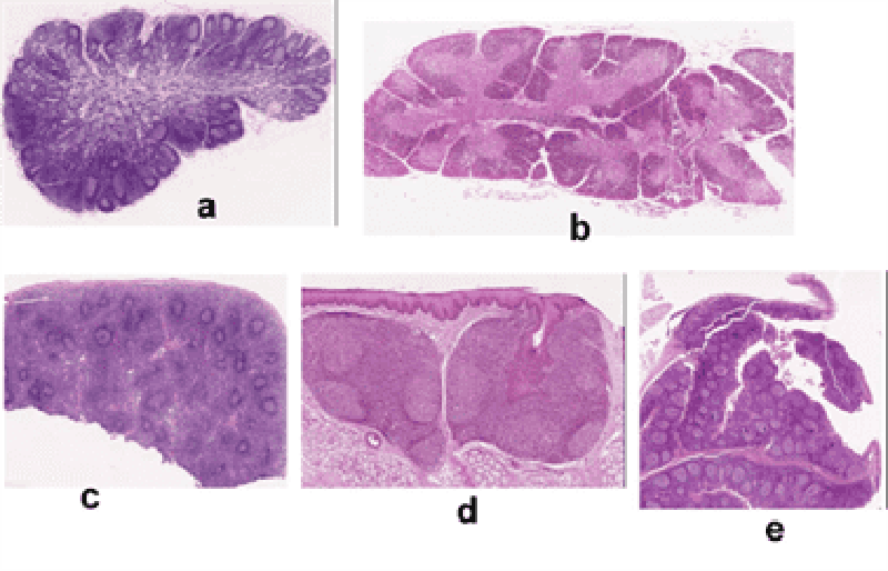
1. Which of the photomicrographs above shows a section of the Thymus?
A
B
C
D
E
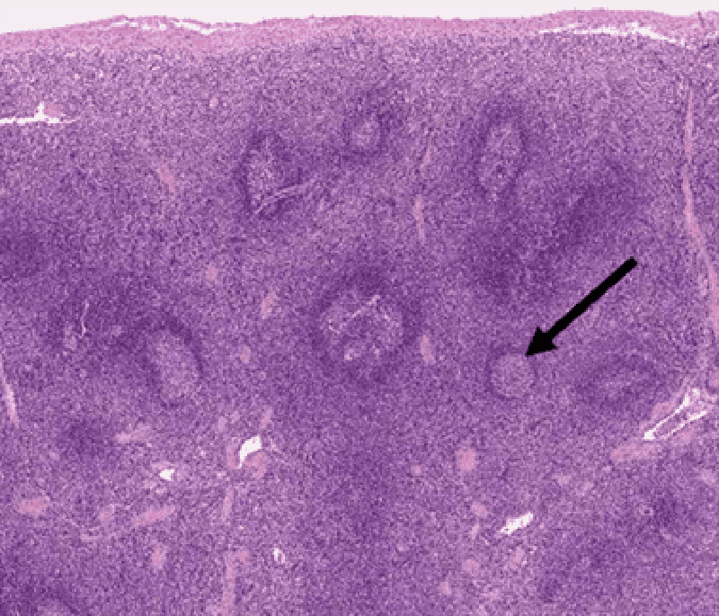
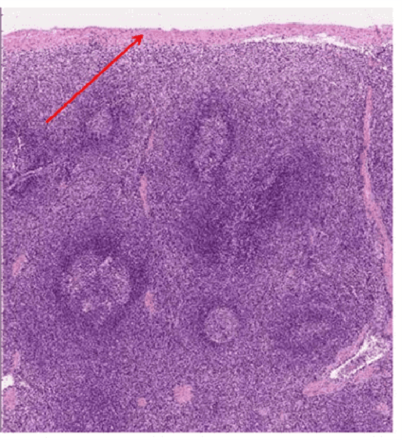
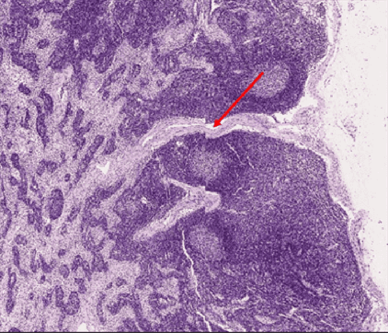
2. Identify area at tip of black arrow.
Red pulp
MALT
Cortex of lymph node
White pulp
Cortex of thymus
3. Tip of red arrow is in lumen of:
Follicular artery
Trabecular artery
Central artery
Penicilar artery
Sheathed artery
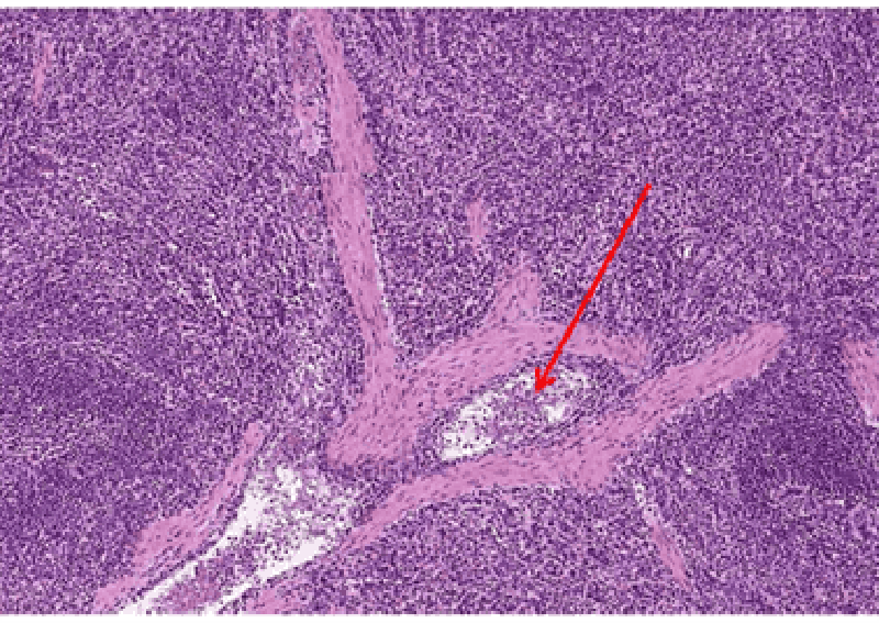
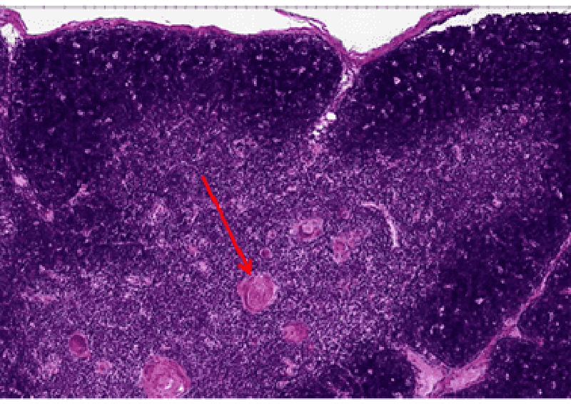
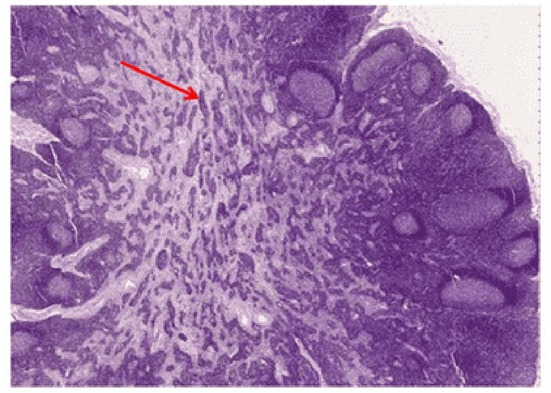
4. Identify structure at tip of red arrow.
Follicular artery
Trabecular artery
Central artery
Penicilar artery
Sheathed artery
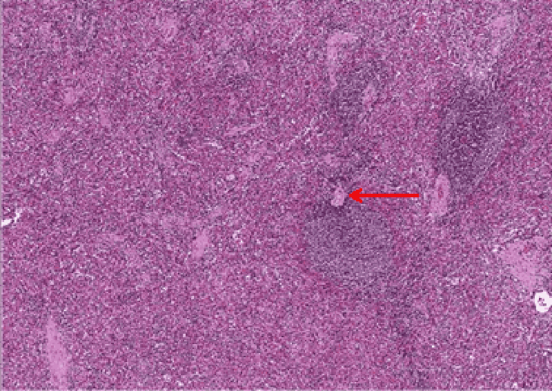
5. Identify structure at tip of red arrow.
Capsule
Keratinized stratified squamous epithelium
Trabecula
Non-keratinized squamous epithelium
Respiratory epithelium
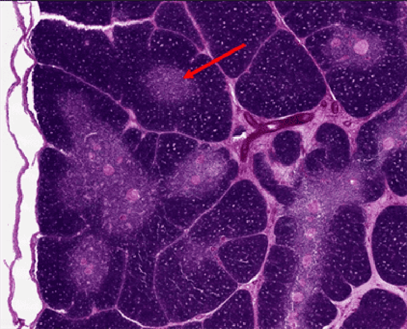
6. Identify structure at tip of red arrow.
Nerve
Central artery
Medullary cord
Trabecular artery
Thymic corpuscle
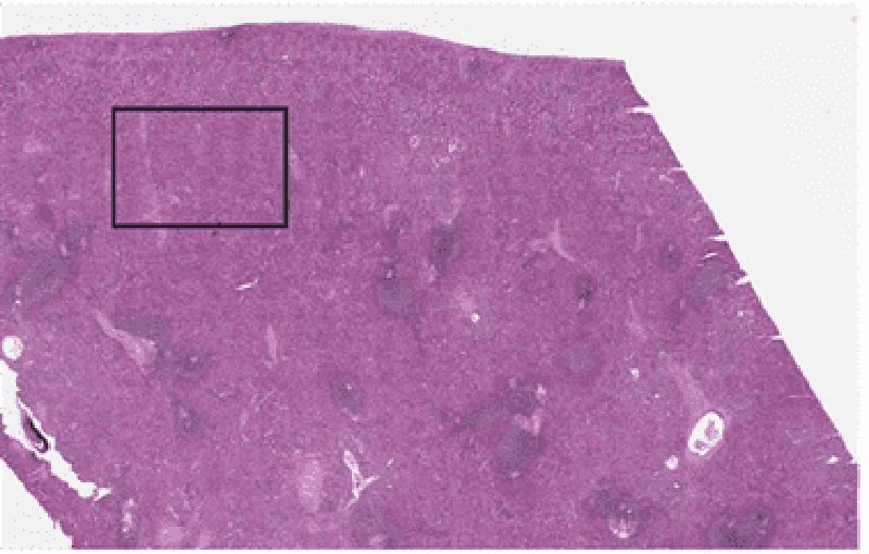
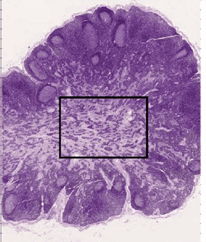
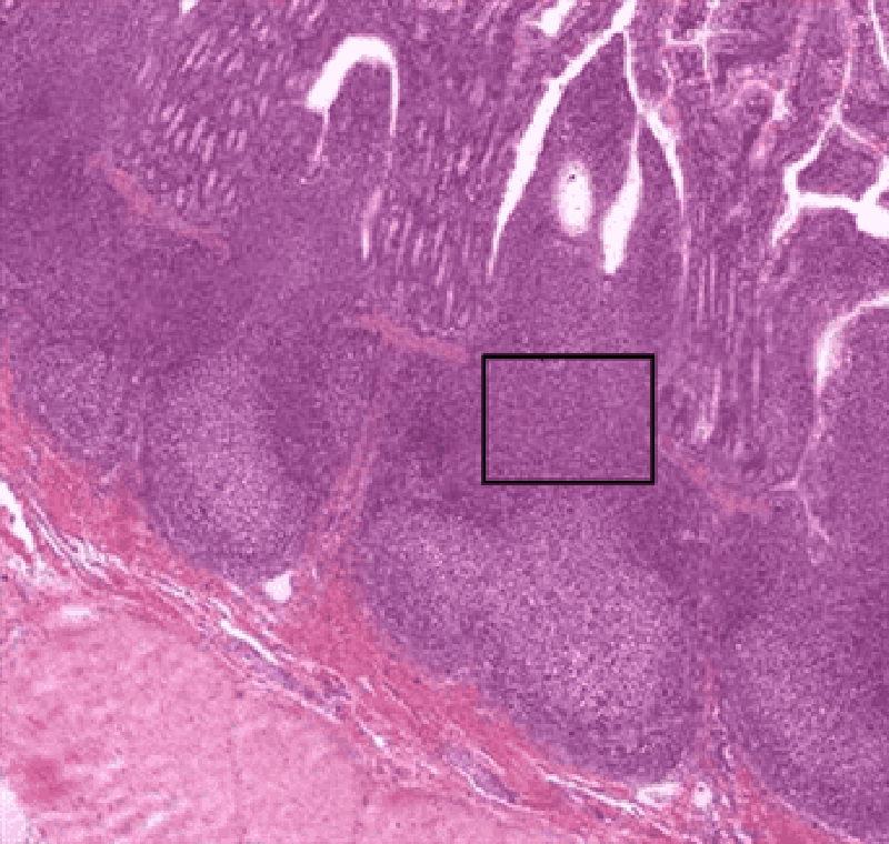
7. Identify region inside black rectangle
White pulp
Cortex
Red pulp
Dense lymphoid tissue
Medulla
8. Identify light staining region at tip of red arrow.
Cortex of thymus
Red pulp of spleen
Medulla of lymph node
Cortex of lymph node
Medulla of thymus
9. Identify area inside black rectangle.
Cortex of lymph node
Red pulp
Cortex of thymus
Medulla of thymus
Medulla of lymph node
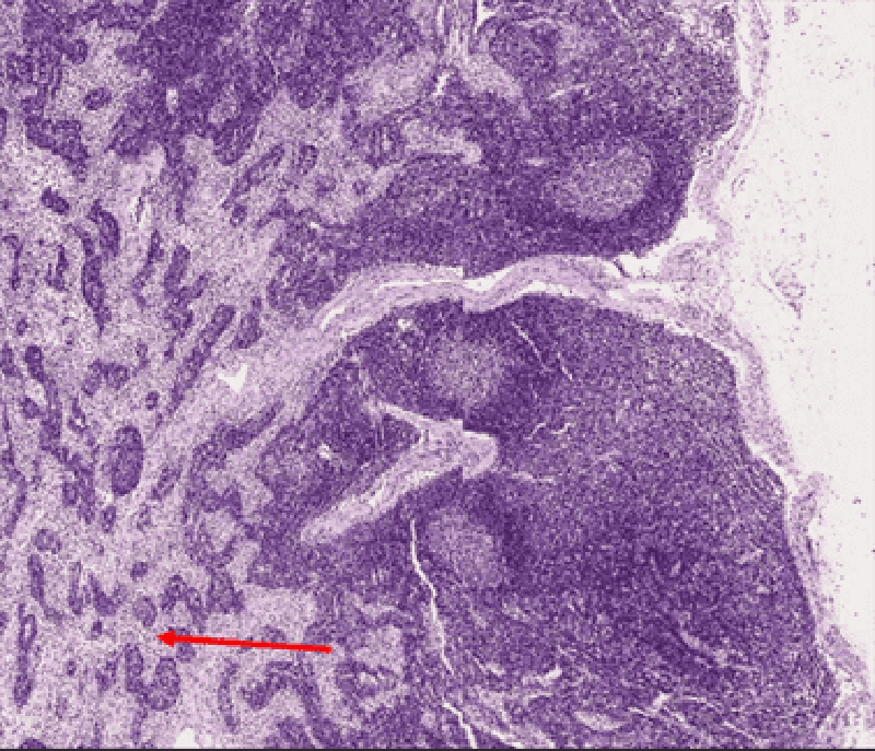
10. Tip of red arrow is in lumen of:
Subcapsular sinus
Tonsilar crypt
Trabecular artery
Trabecular sinus
Medullary sinus
11. Tip of red arrow is in lumen of:
Trabecular artery
Trabecular sinus
Subscapular sinus
Medullary sinus
Tonsilar crypt

12. Which of the photomicrographs above shows a section of the lingual tonsil?
A
B
C
D
E
13. Identify structure at tip of red arrow.
Splenic cord of Billroth
Trabecula
Medullary cord
Lymphoid nodule
White pulp

14. Which of the photomicrographs above shows a section of the palatine tonsil?
A
B
C
D
E

15. Which of the photomicrographs above shows a section of the Spleen?
A
B
C
D
E

16. Which of the photomicrographs above shows a section of the lymph node?
A
B
C
D
E
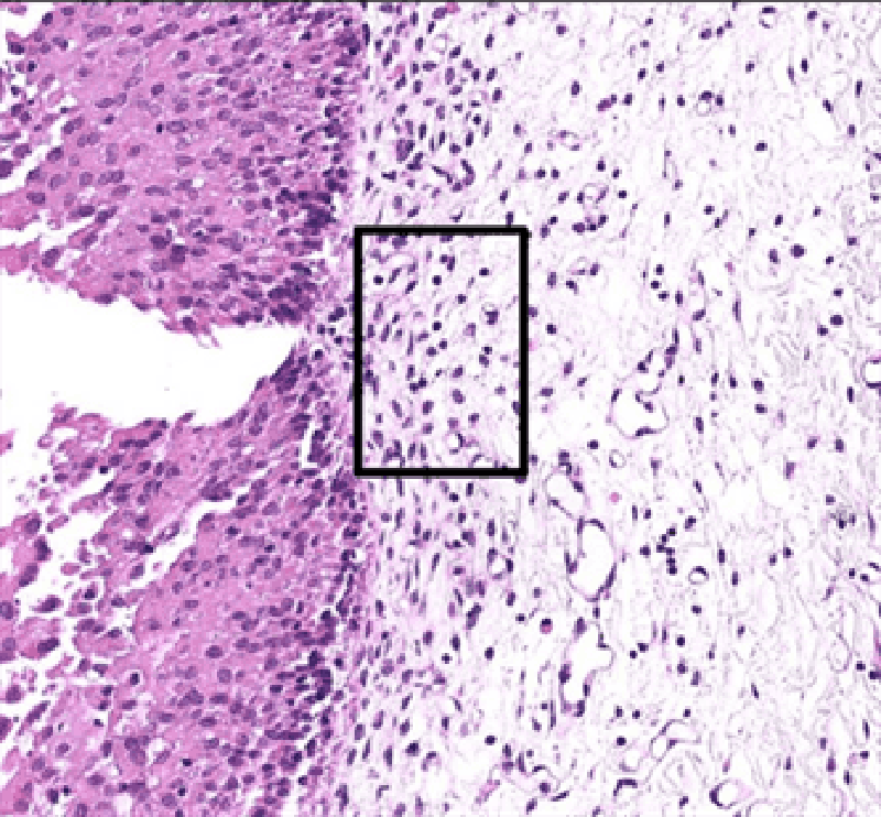
17. Identify tissue inside black rectangle.
PALS
Secondary lymphoid nodule
Loose lymphoid tissue
Primary lymphoid tissue
Dense lymphoid tissue
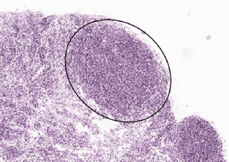
18. Identify structure inside black oval.
Dense lymphoid tissue
PALS
Primary lymphoid tissue
Secondary lymphoid tissue
Loose lymphoid tissue
19. Identify tissue inside black rectangle.
Primary lymphoid tissue
Secondary lymphoid tissue
Dense lymphoid tissue
Loose lymphoid tissue
Dense regular connective tissue
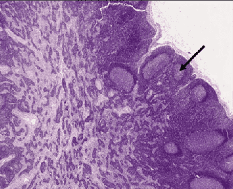
20. Identify region at tip of black arrow.
Germinal center
Primary lymphoid tissue
Dense lymphoid tissue
Corona
White pulp
{"name":"Lymph Module - Histo Pracs", "url":"https://www.quiz-maker.com/QPREVIEW","txt":"Test your knowledge on the intricate structures of lymphatic tissues and organs with our comprehensive 20-question quiz. Perfect for students and professionals in the field of histology, this quiz presents various photomicrographs and challenges you to identify key components related to the lymphatic system.Each question is thoughtfully crafted to enhance your understanding of:The thymus and spleen structureLymph nodes and tonsilsMajor lymphatic tissues","img":"https:/images/course2.png"}
More Quizzes
Histo retake on entry cycle 1
221171
Histology 2E
29140
Fdv
100
Nursing MCQ
211030
Scarlett Johansson: How Obsessed Are You?
201017776
Grade 12 English Practice Exam - Free Final Review
201016996
Pediatric Nursing 3.0 Test - Free NCLEX Practice
201021775
Cups and Saucers - Free Tea Party Etiquette
201022654
A Loose Union of Independent States - Free Civics
201021657
Hard IQ Test - Free Online Challenge: Can You Score High?
201021775
Computer Network - Test Your Networking Knowledge
201017571
What Season Am I - Spring, Summer, Fall or Winter?
201016583