
Cell biology quiz group 11

Cell Biology Challenge
Test your knowledge on cell biology with this comprehensive quiz designed for students and enthusiasts alike. With 40 thoughtfully crafted questions, you'll explore various aspects of cell structures, fun
Whether you are preparing for exams or simply eager to learn more about the fascinating world of cells, this quiz is an excellent resource!
- 40 Questions
- Focus on histology and cell fun
ction - Multichoice format for easy answering
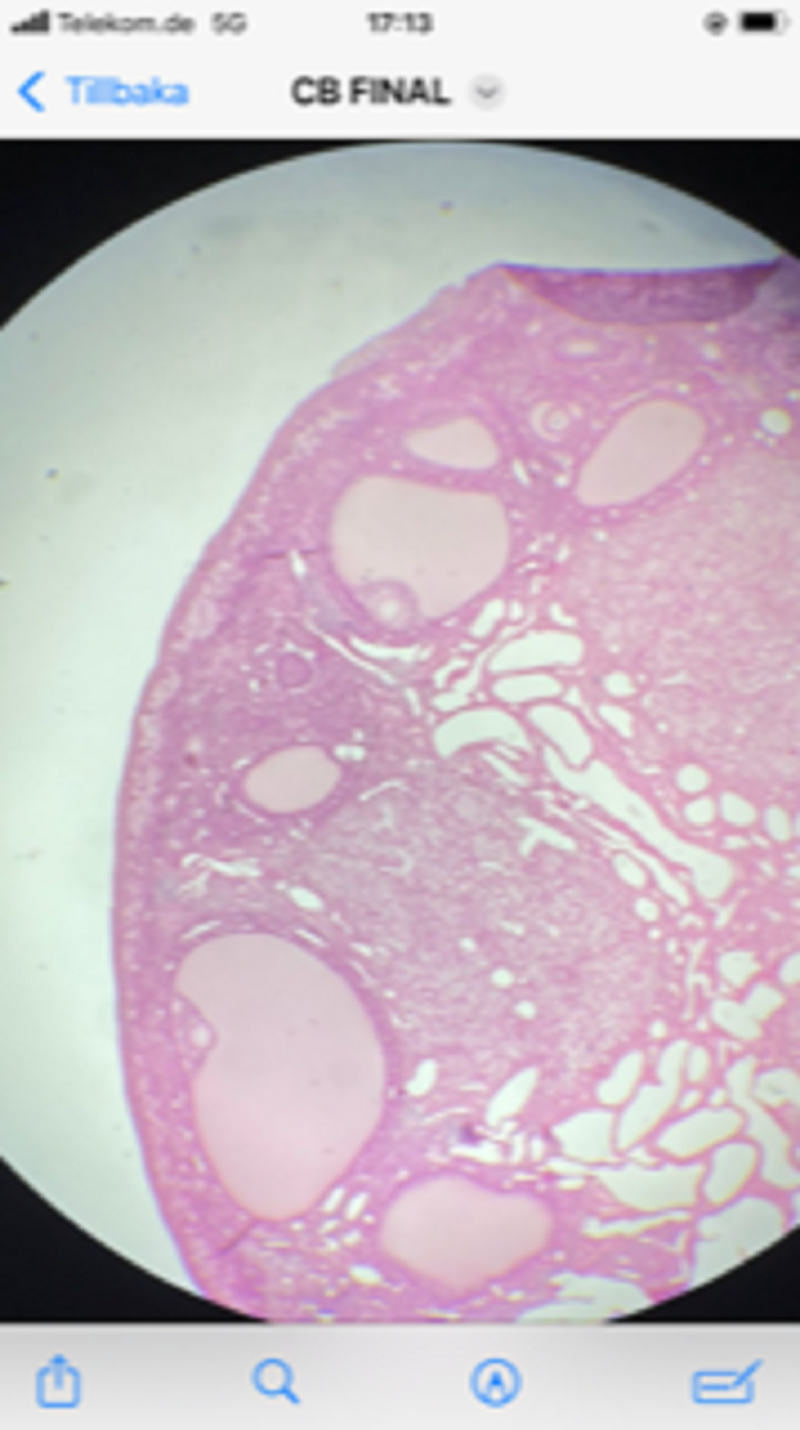
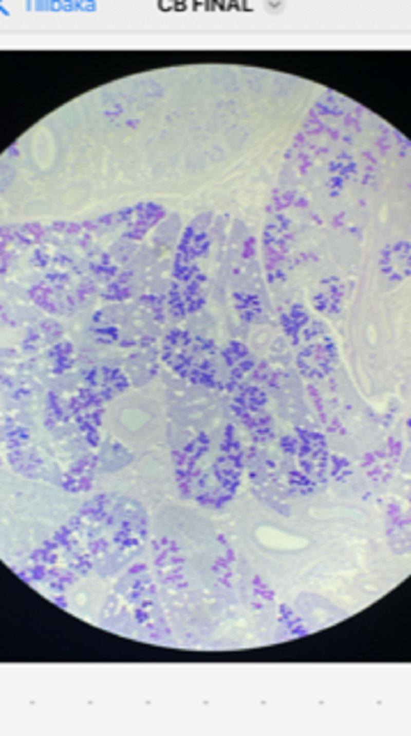
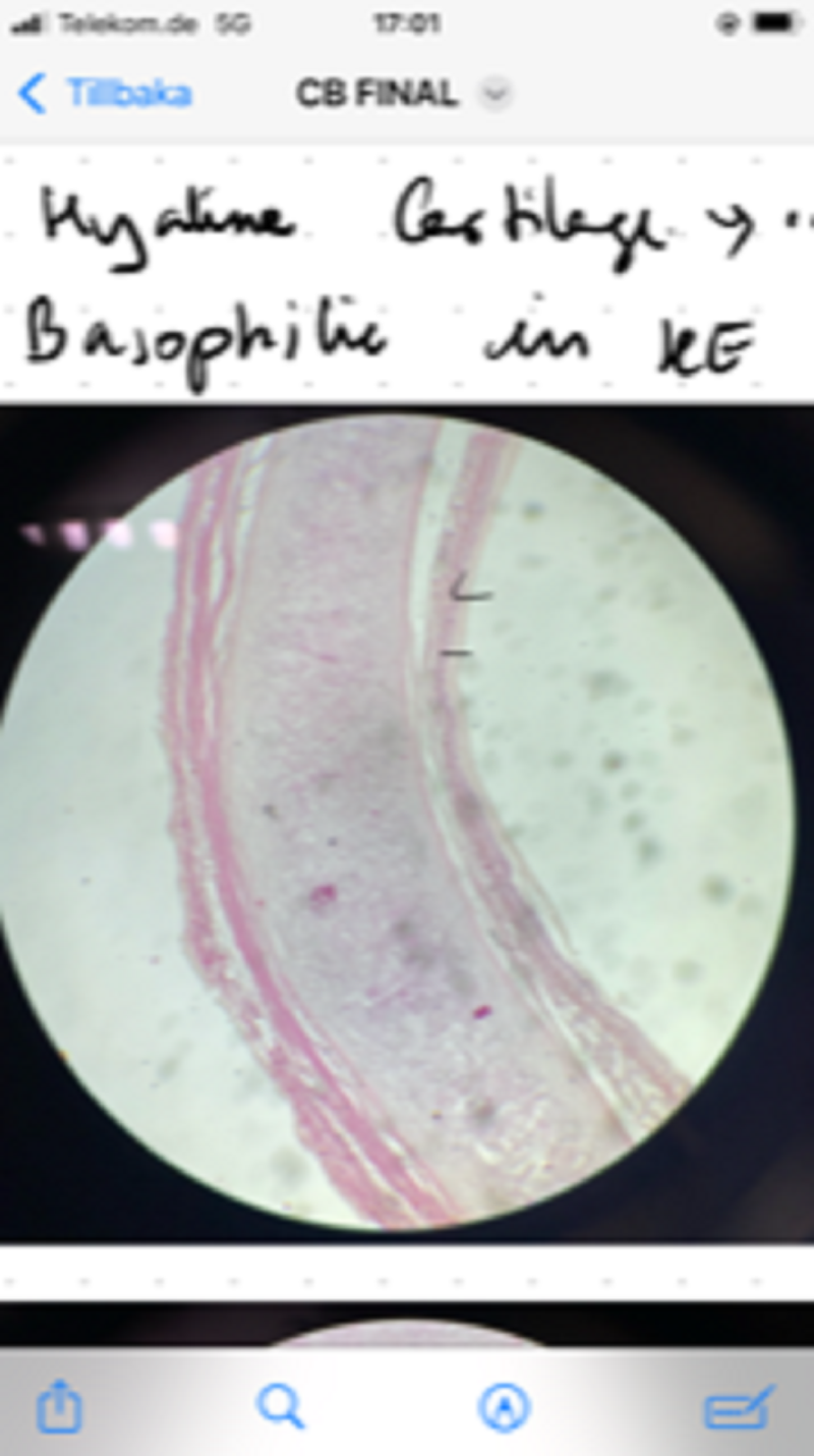
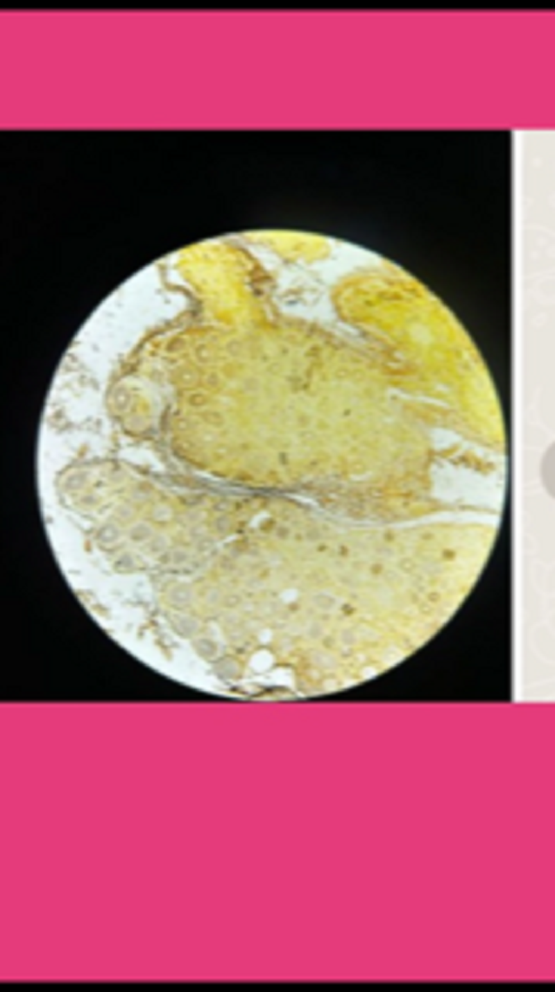
Meiosis. Oogenesis. Ovary. HE stain
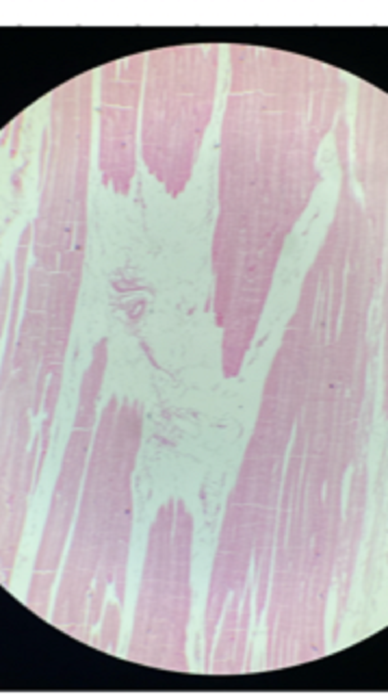
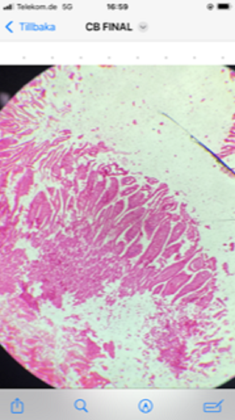
Syncytium. Striated muscle fibres. Skeletal muscle. H&E Stain
Kinocilia. Epithelial cells. Trachea. HE stain
Collagen fibers. Skin. HE stain
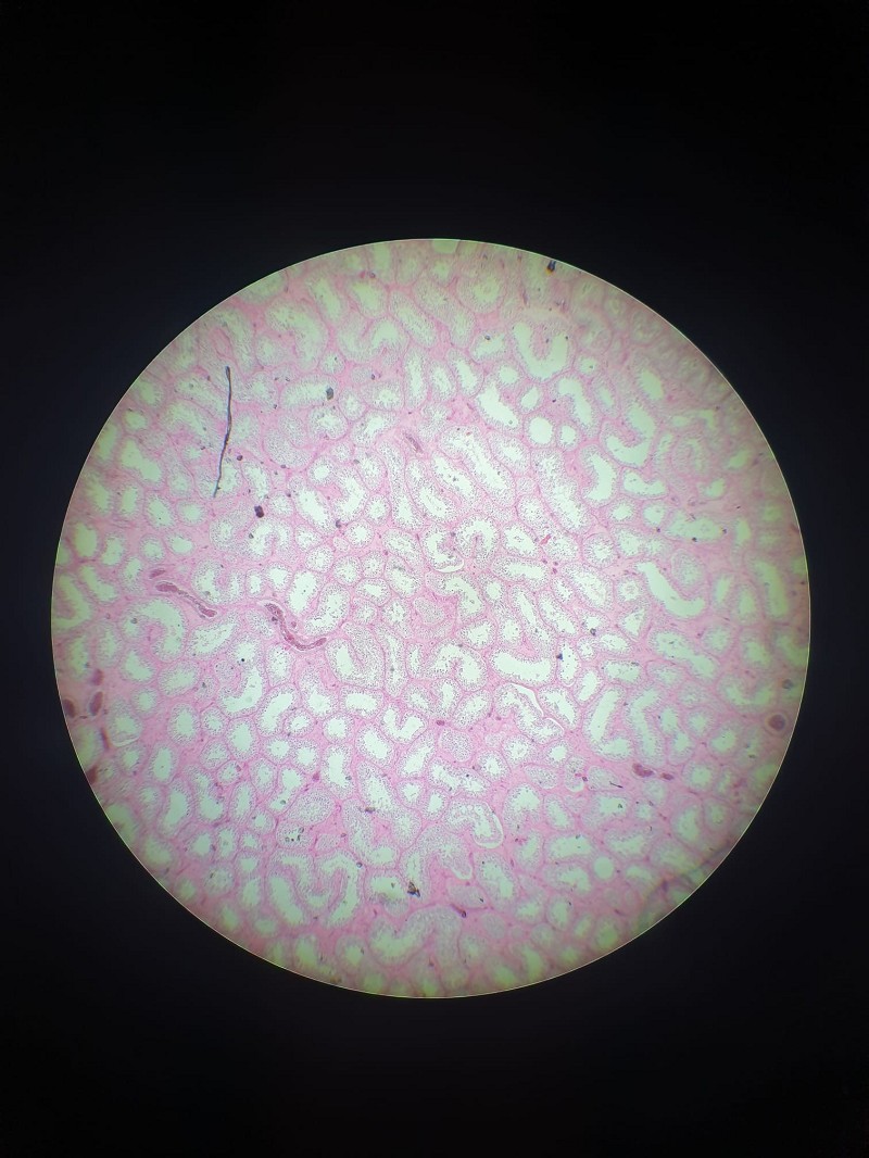
Microvilli. Brush border. Nephrocytes. Kidney. HE stain
Syncytium. Striated muscle fibres. Skeletal muscle. H&E Stain
Fusiform cells. Fibroblasts. Gingival tissue. Mandible. H&E Stain
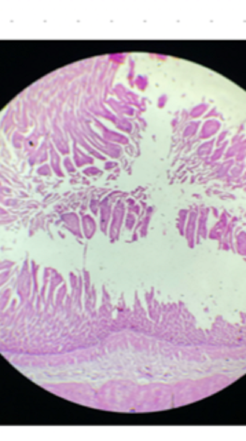
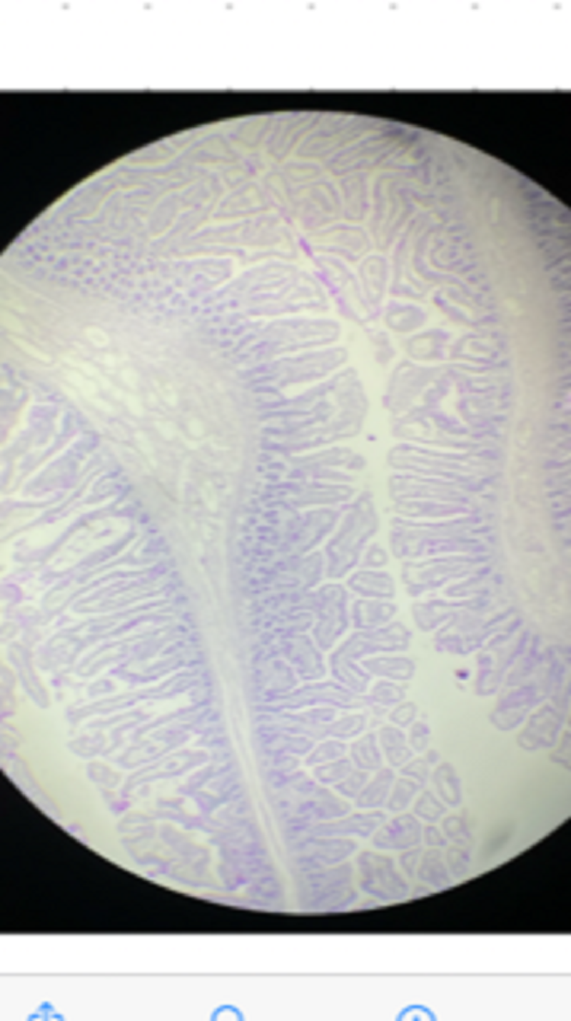
Columnar cells. Enterocytes. Duodenum. H&E Stain
Columnar cells. Enterocytes. Duodenum. H&E Stain
Neutral & acid mucopolysaccharides. Secreting cells. Salivary gland. PAS + AB stain
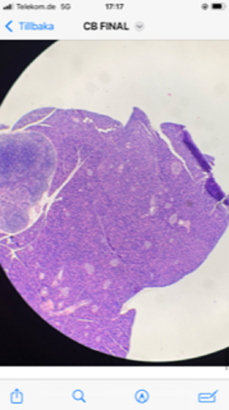
RER. Acinar cells. Endocrine secretion. Islets of Langerhans. Pancreas. HE stain
Kinocilia. Epithelial cells. Trachea. HE stain
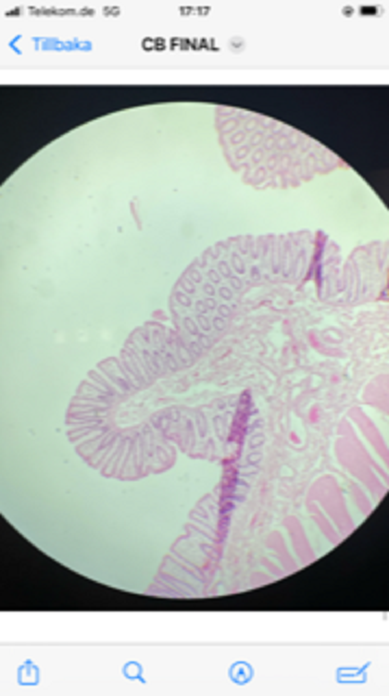
Apocrine secretion. Goblet cells. Colon. HE stain
Fusiform cells. Fibroblasts. Gingival tissue. Mandible. H&E Stain
Columnar cells. Enterocytes. Duodenum. H&E Stain
Collagen fibers. Skin. HE stain
Neutral mucopolysaccharides. Goblet cells. Small intestine. PAS + H stain
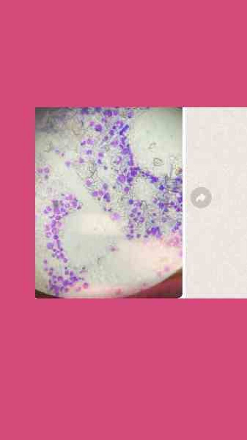
Polygonal cells. Cervical (Pap) smear. May GrünwaldGiemsa (MGG) Stain
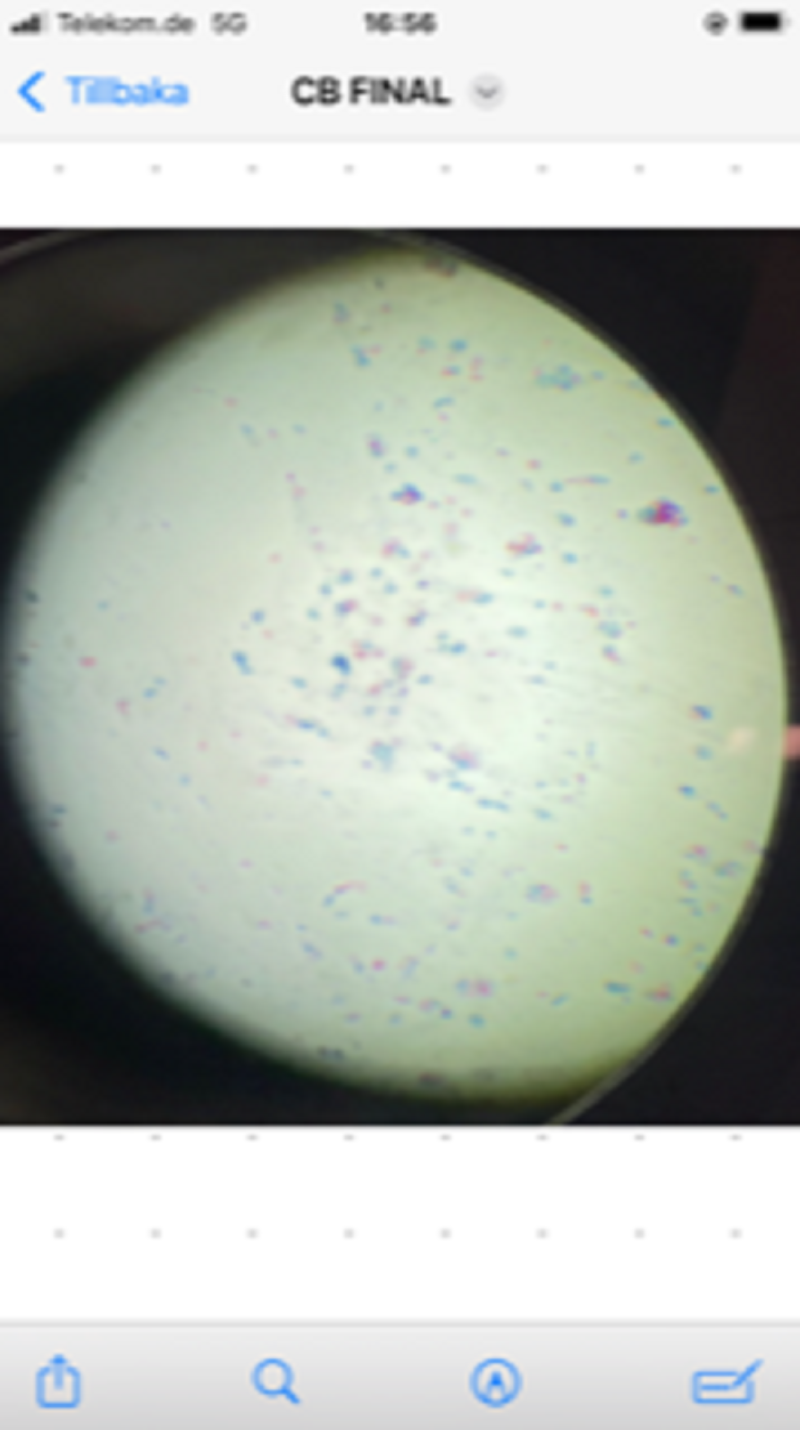
Segmented nucleus (neutrophil), bilobed nucleus (eosinophil), bi (tri ) - lobed nucleus (basophil), round nucleus (lymphocyte), reniform nucleus (monocyte). Peripheral blood smear. May Grünwald Giemsa (MGG) stain
Kinocilia. Epithelial cells. Trachea. HE stain
Merocrine secretion. Serous, mucous and mixed acini. Salivary gland. HE stain
Apocrine secretion. Goblet cells. Colon. HE stain
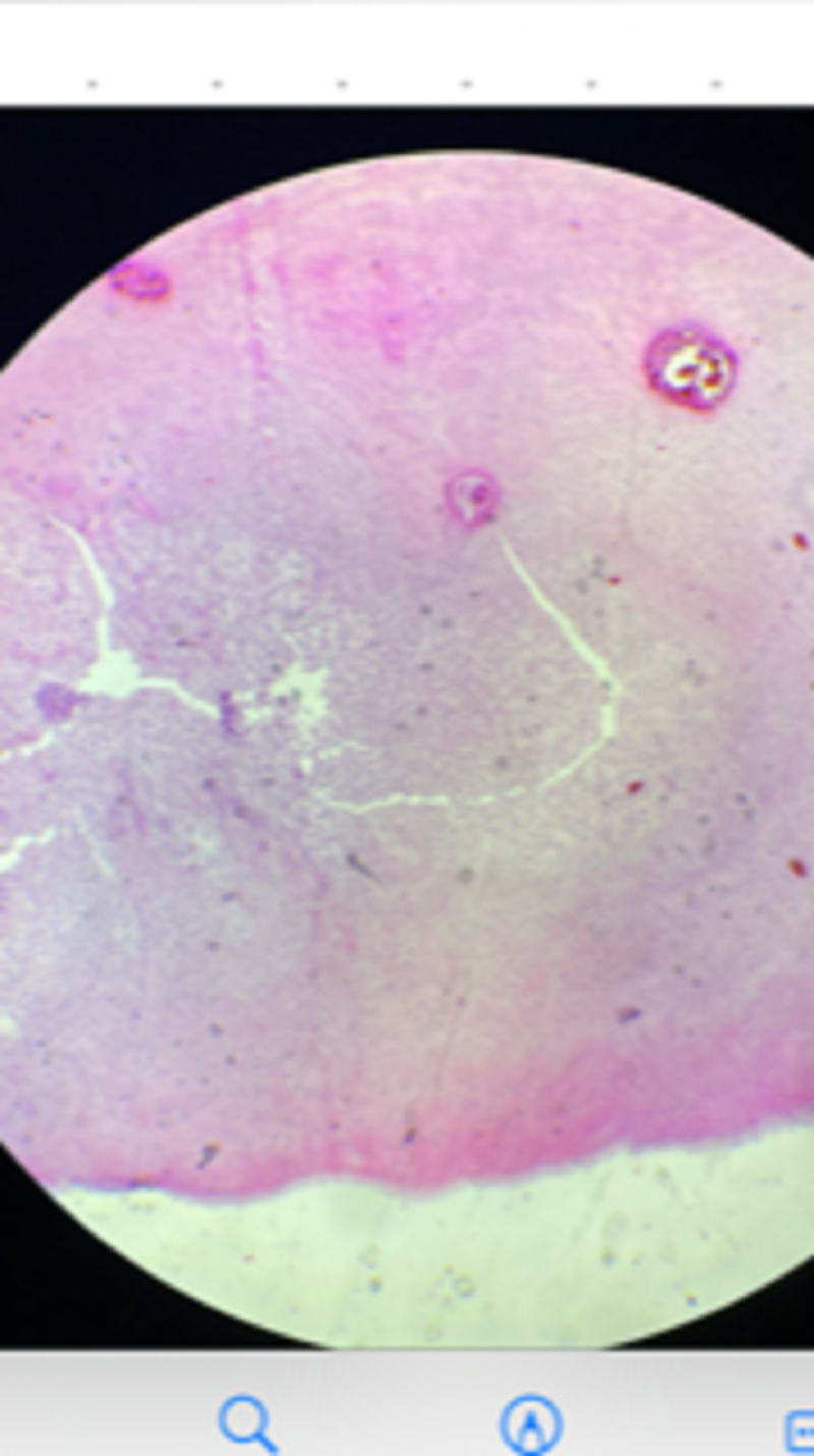
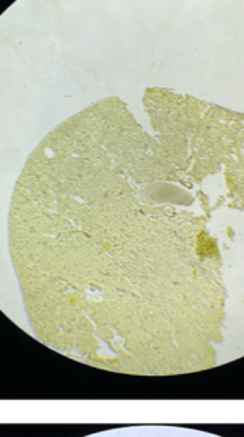
Amitosis. Hepatocytes. Berg Bodies. Liver HE stain
Lobulated nucleus .Endomitosis Megakaryocyte. Bone marrow smear. MGG stain
Lobulated nucleus .Endomitosis Megakaryocyte. Bone marrow smear. MGG stain
Basement membrane. Nephrocytes. Renal tubules. Kidney. PAS stain
Meiosis. Oogenesis. Ovary. HE stain
Apocrine secretion. Goblet cells. Colon. HE stain
Merocrine secretion. Serous, mucous and mixed acini. Salivary gland. HE stain
Amitosis. Hepatocytes. Berg Bodies. Liver HE stain
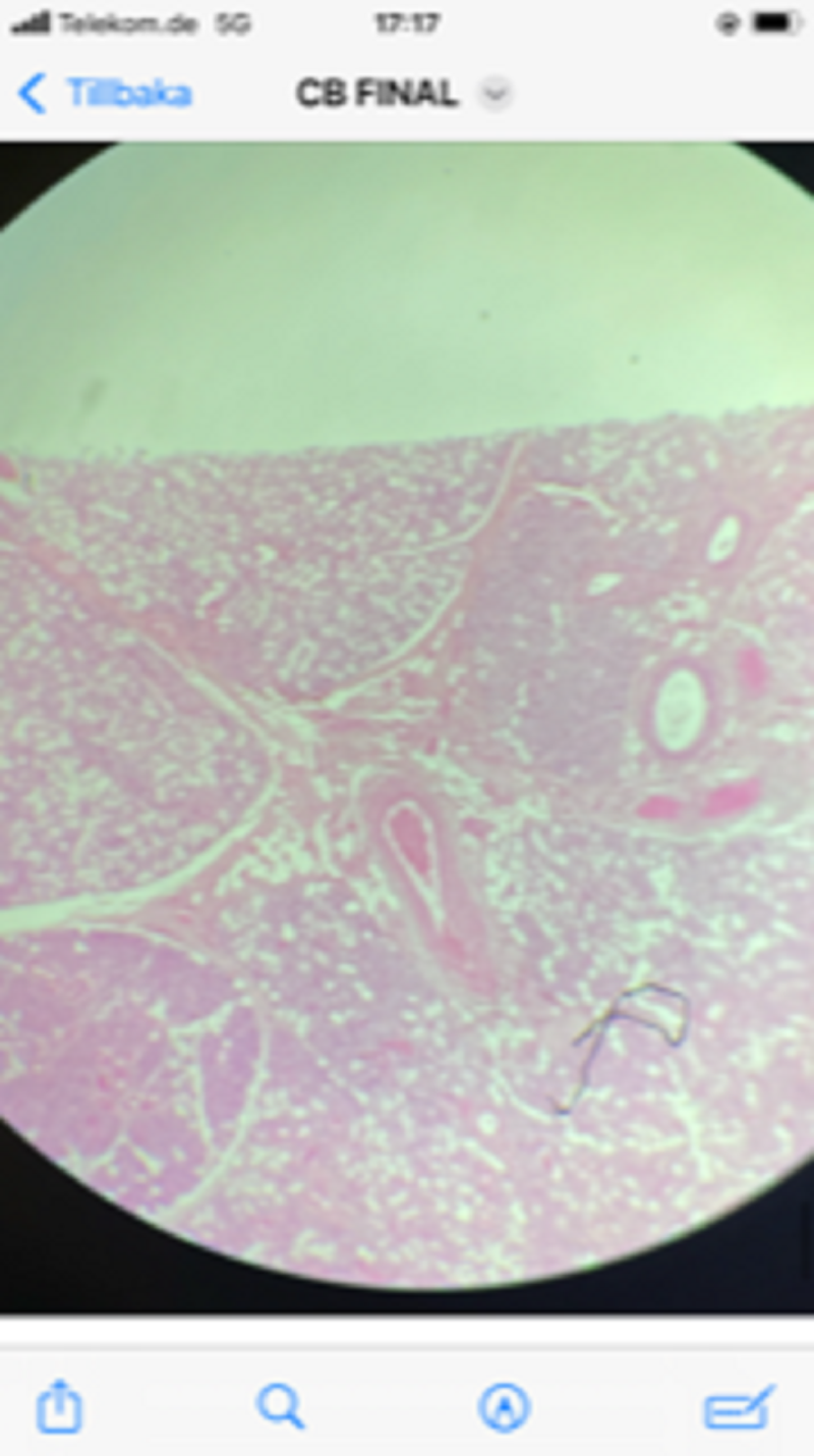
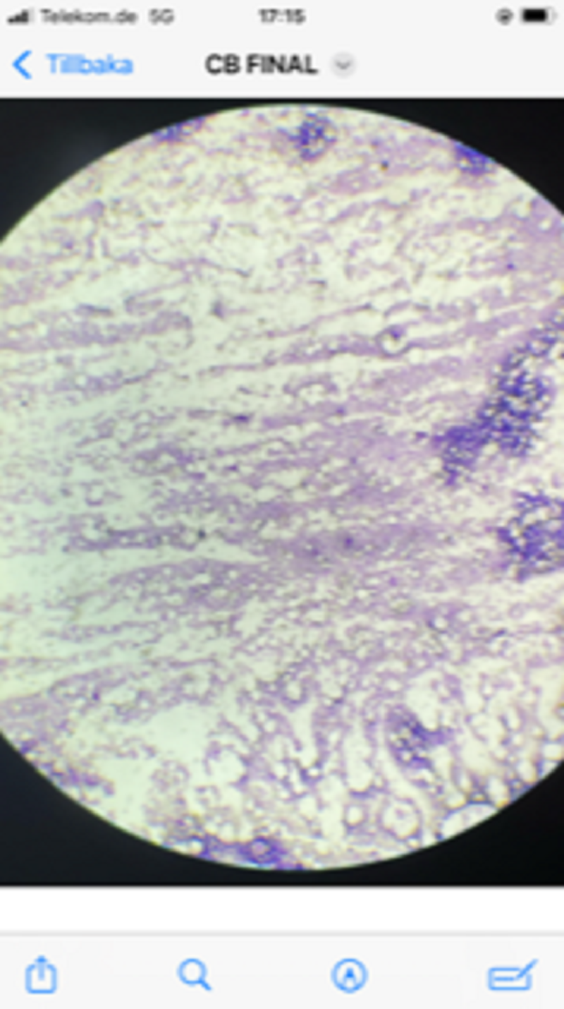
Meiosis. Spermatogenesis. Testis. HE stain
Reticulin fibres. Basement membrane. Nephrocytes. Renal tubules. Kidney. Silver impregnation
Fusiform cells. Fibroblasts. Gingival tissue. Mandible. H&E Stain
Syncytium. Striated muscle fibres. Skeletal muscle. H&E Stain
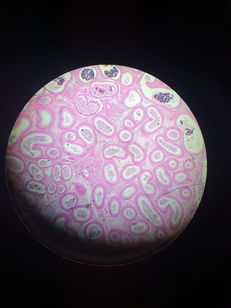
Stereocilia. Epithelial cells. Epididymis. HE stain
Collagen fibers. Skin. HE stain
RER. Nissl Bodies. Neurons. Spinal cord. Toluidine Blue Stain
Amitosis. Hepatocytes. Berg Bodies. Liver HE stain
RER. Acinar cells. Endocrine secretion. Islets of Langerhans. Pancreas. HE stain
Merocrine secretion. Serous, mucous and mixed acini. Salivary gland. HE stain
NOT 100% sure about this one, please correct me if its wrong! <3
Merocrine secretion. Serous, mucous and mixed acini. Salivary gland. HE stain
Apocrine secretion. Goblet cells. Colon. HE stain
RER. Acinar cells. Endocrine secretion. Islets of Langerhans. Pancreas. HE stain
Lobulated nucleus .Endomitosis. Megakaryocyte. Bone marrow smear. MGG stain
Neutral mucopolysaccharides. Goblet cells. Small intestine. PAS + H stain
Acid mucopolysaccharides. Goblet cells. Colon. Alcian blue (AB) stain
Glycogen. Hepatocytes. Liver. Periodic Acid Schiff (PAS) stain
Neutral & acid mucopolysaccharides. Secreting cells. Salivary gland. PAS + AB stain
Stellate cells. Motor neurons. Spinal cord. H&E Stain
Cilia and microvilli. Ependymal cells. Central canal. Spinal cord. HE stain
Glycogen. Hepatocytes. Liver. Periodic Acid Schiff (PAS) stain
Mitosis. Bone marrow smear. MGG stain
Basement membrane. Nephrocytes. Renal tubules. Kidney. PAS stain
Cilia and microvilli. Ependymal cells. Central canal. Spinal cord. HE stain
Polygonal cells. Cervical (Pap) smear. May GrünwaldGiemsa (MGG) Stain
Stellate cells. Motor neurons. Spinal cord. H&E Stain
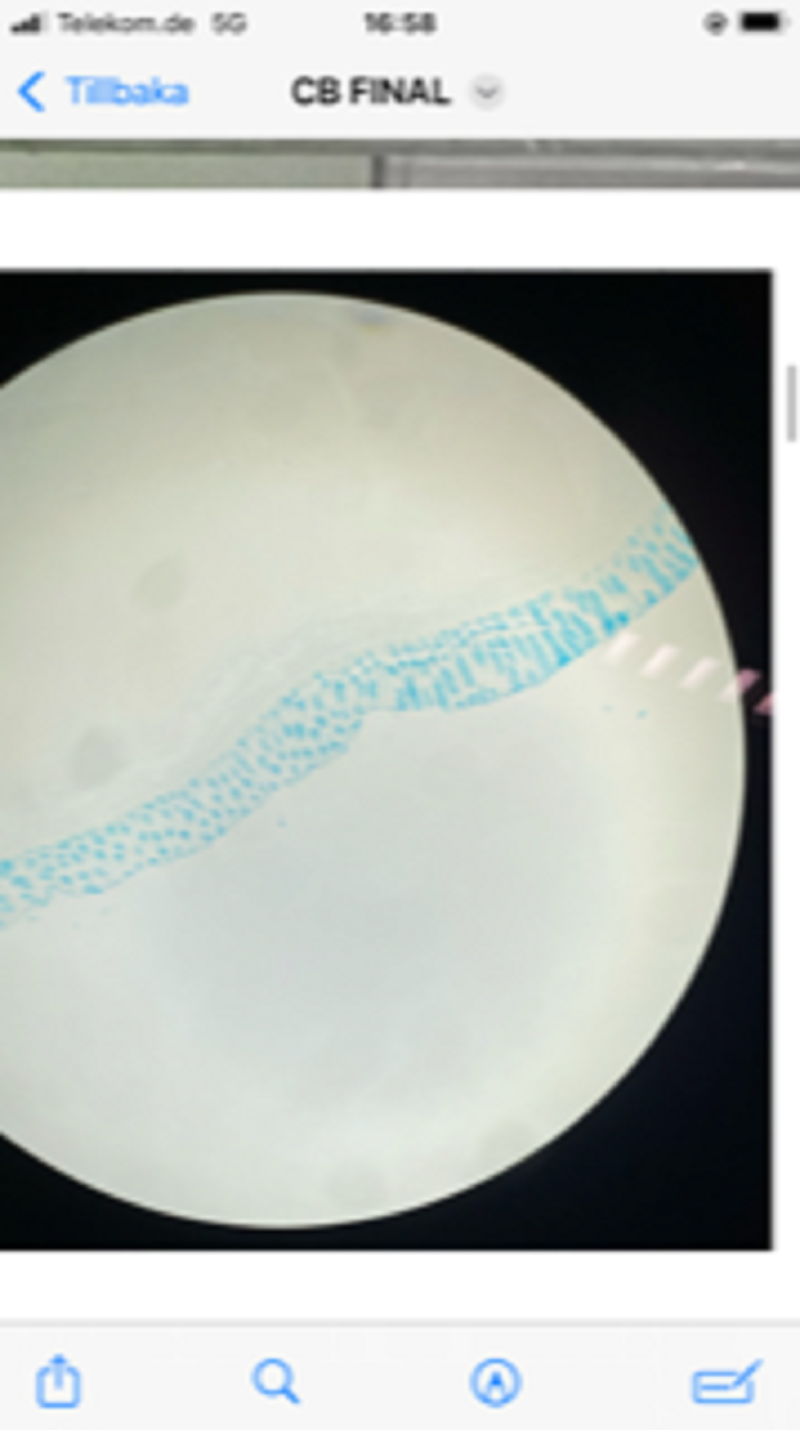
Phospholipids. Artery. Sudan V (Black) stain
Phospholipids. Artery. Sudan VI (Black) stain
Phospholipids. Artery. Sudan IV (Black) stain
Phospholipids. Artery. Sudan IV (Brown) stain
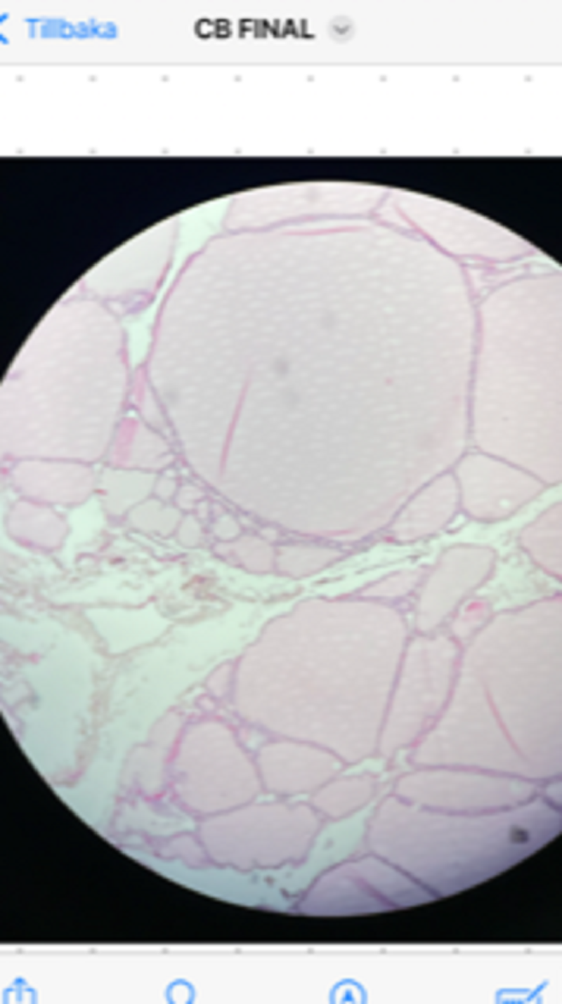
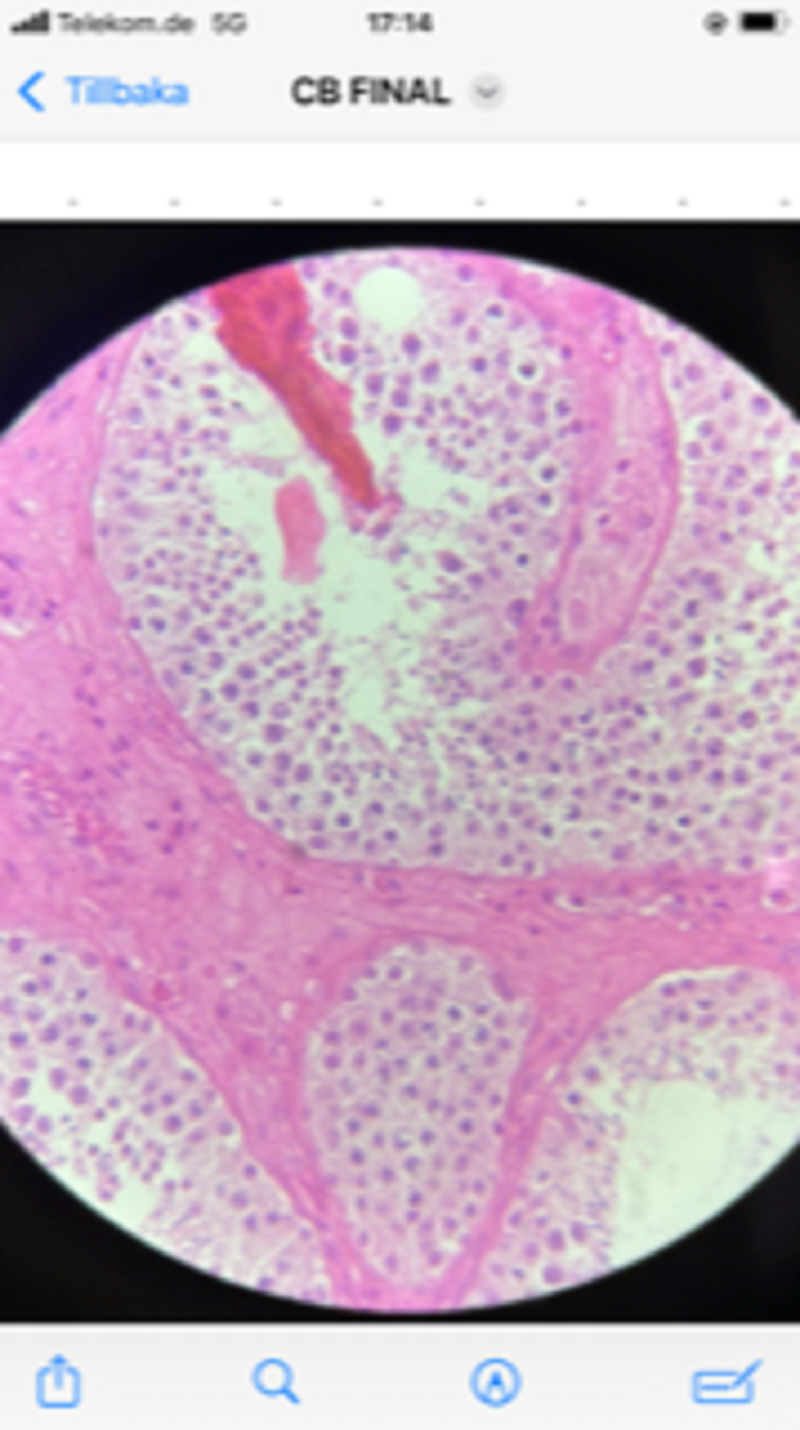
Cuboidal cells. Thyroid follicles. Endocrine secretion. Thyroid. H&E Stain
Stereocilia. Epithelial cells. Epididymis. HE stain
Reticulin fibres. Basement membrane. Nephrocytes. Renal tubules. Kidney. Silver impregnation
Golgi apparatus. Secretory neurons. Hypothalamus. Silver impregnation
Triglycerides. Artery. Sudan III stain
Elastic fibres. Artery cross section. Resorcin fuchsin stain
Polygonal cells. Cervical (Pap) smear. May GrünwaldGiemsa (MGG) Stain
Kinocilia. Epithelial cells. Trachea. HE stain
Reticulin fibres. Basement membrane. Nephrocytes. Renal tubules. Kidney. Silver impregnation
Golgi apparatus. Secretory neurons. Hypothalamus. Silver impregnation
Mitosis. Bone marrow smear. MGG stain
Meiosis. Oogenesis. Ovary. HE stain
Stellate cells. Motor neurons. Spinal cord. H&E Stain
Cilia and microvilli. Ependymal cells. Central canal. Spinal cord. HE stain
Basement membrane. Nephrocytes. Renal tubules. Kidney. PAS stain
RER. Nissl Bodies. Neurons. Spinal cord. Toluidine Blue Stain
Stellate cells. Motor neurons. Spinal cord. H&E Stain
Fusiform cells. Fibroblasts. Gingival tissue. Mandible. H&E Stain
Glycogen. Hepatocytes. Liver. Periodic Acid Schiff (PAS) stain
Segmented nucleus (neutrophil), bilobed nucleus (eosinophil), bi (tri ) - lobed nucleus (basophil), round nucleus (lymphocyte), reniform nucleus (monocyte). Peripheral blood smear. May Grünwald Giemsa (MGG) stain
Phospholipids. Artery. Sudan IV (Black) stain
Kinocilia. Epithelial cells. Trachea. HE stain
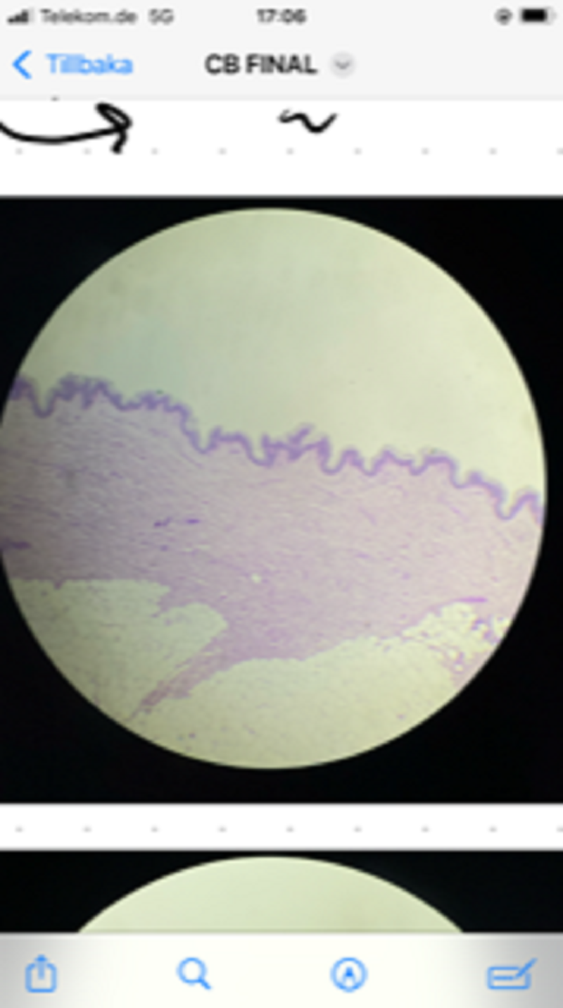
Basement membrane. Skin. PAS stain
Elastic fibres. Artery cross section. Resorcin fuchsin stain
Cilia and microvilli. Ependymal cells. Central canal. Spinal cord. HE stain
Flattened nucleus. Adipocyte. Adipose tissue. Skin. HE stain
Mitosis. Bone marrow smear. MGG stain
Golgi apparatus. Secretory neurons. Hypothalamus. Silver impregnation
Cuboidal cells. Thyroid follicles. Endocrine secretion. Thyroid. H&E Stain
Microvilli. Brush border. Nephrocytes. Kidney. HE stain
Stereocilia. Epithelial cells. Epididymis. HE stain
Meiosis. Spermatogenesis. Testis. HE stain
Columnar cells. Enterocytes. Duodenum. H&E Stain
Fusiform cells. Fibroblasts. Gingival tissue. Mandible. H&E Stain
Microvilli. Brush border. Nephrocytes. Kidney. HE stain
Amitosis. Hepatocytes. Berg Bodies. Liver HE stain
Syncytium. Striated muscle fibres. Skeletal muscle. H&E Stain
Fusiform cells. Fibroblasts. Gingival tissue. Mandible. H&E Stain
Collagen fibers. Skin. HE stain
Columnar cells. Enterocytes. Duodenum. H&E Stain
Columnar cells. Enterocytes. Duodenum. H&E Stain
Cuboidal cells. Thyroid follicles. Endocrine secretion. Thyroid. H&E Stain
Neutral mucopolysaccharides. Goblet cells. Small intestine. PAS + H stain
Lobulated nucleus .Endomitosis Megakaryocyte. Bone marrow smear. MGG stain
Neutral & acid mucopolysaccharides. Secreting cells. Salivary gland. PAS + AB stain
RER. Nissl Bodies. Neurons. Spinal cord. Toluidine Blue Stain
Acid mucopolysaccharides. Goblet cells. Colon. Alcian blue (AB) stain
Glycogen. Hepatocytes. Liver. Periodic Acid Schiff (PAS) stain
NOT 100% sure about this one, please correct me if Im wrong. <3
Syncytium. Striated muscle fibres. Skeletal muscle. H&E Stain
Fusiform cells. Fibroblasts. Gingival tissue. Mandible. H&E Stain
Meiosis. Oogenesis. Ovary. HE stain
Collagen fibers. Skin. HE stain
RER. Nissl Bodies. Neurons. Spinal cord. Toluidine Blue Stain
Amitosis. Hepatocytes. Berg Bodies. Liver HE stain
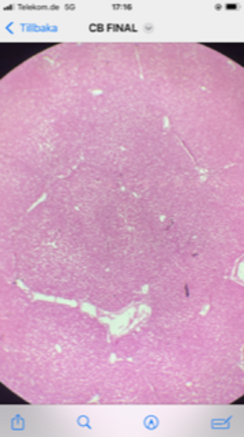
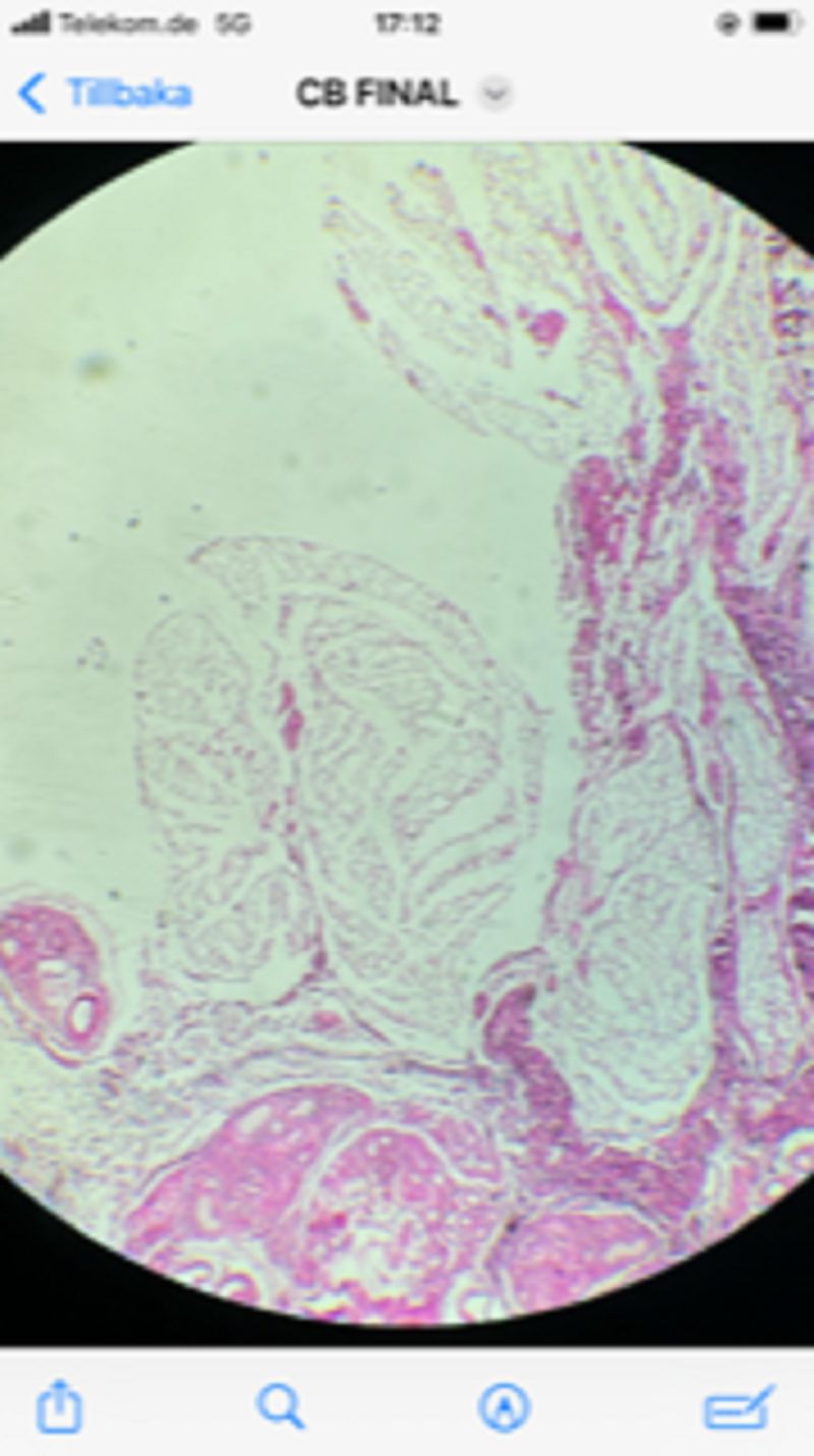
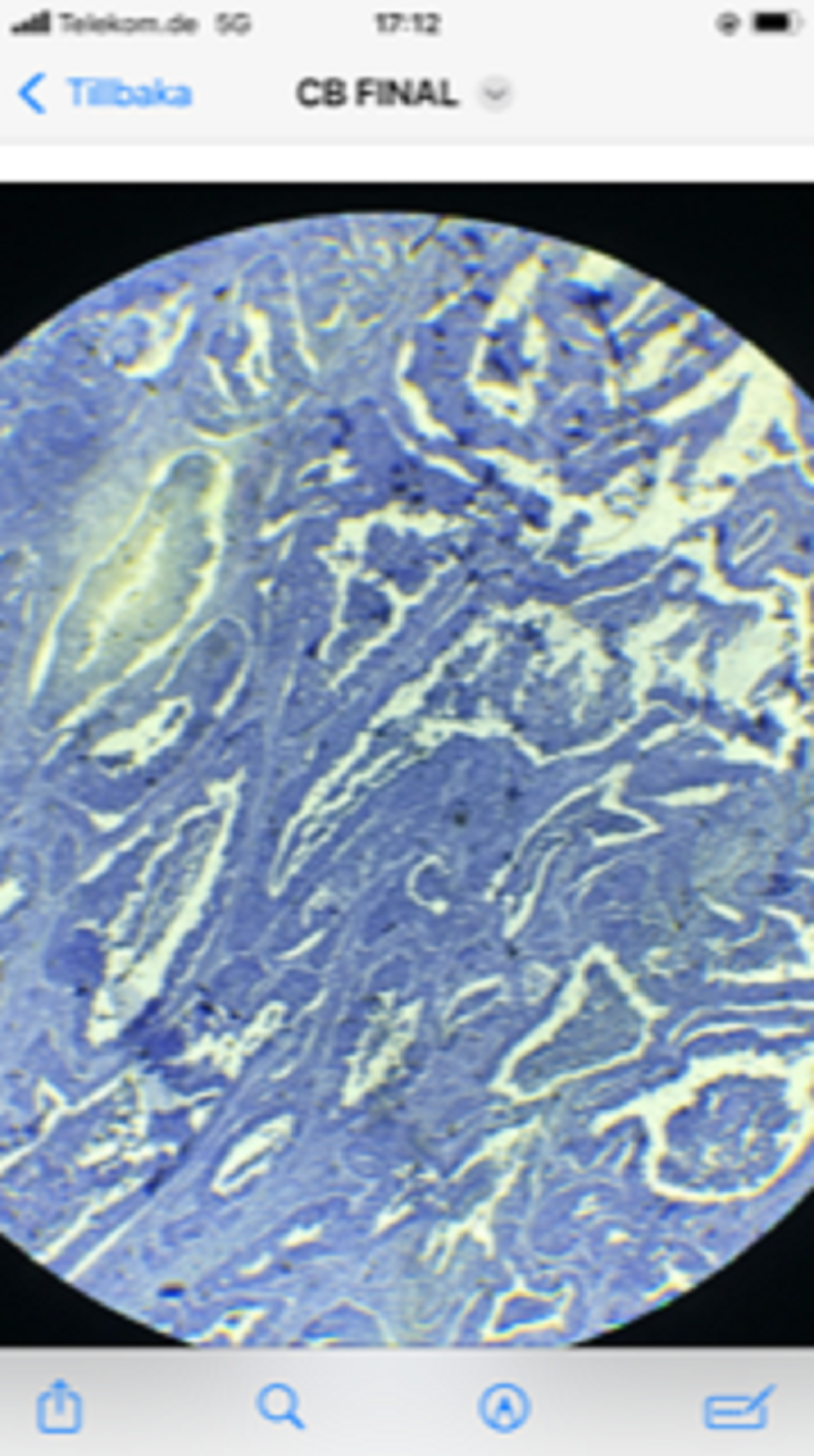
Monstruous, irregular nuclei. Neoplastic cells. Malignant tumor. Iron/ferric hematoxylin (FeH) stain
Neutral & acid mucopolysaccharides. Secreting cells. Salivary gland. PAS + AB stain
Neutral & acid mucopolysaccharides. Secreting cells. Salivary gland. PAS + AB stain
RER. Acinar cells. Endocrine secretion. Islets of Langerhans. Pancreas. HE stain
Lobulated nucleus .Endomitosis. Megakaryocyte. Bone marrow smear. MGG stain
Polygonal cells. Cervical (Pap) smear. May GrünwaldGiemsa (MGG) Stain
Segmented nucleus (neutrophil), bilobed nucleus (eosinophil), bi (tri ) - lobed nucleus (basophil), round nucleus (lymphocyte), reniform nucleus (monocyte). Peripheral blood smear. May Grünwald Giemsa (MGG) stain
Amitosis. Hepatocytes. Berg Bodies. Liver HE stain
RER. Acinar cells. Endocrine secretion. Islets of Langerhans. Pancreas. HE stain
Merocrine secretion. Serous, mucous and mixed acini. Salivary gland. HE stain

Triglycerides. Artery. Sudan III stain
Phospholipids. Artery. Sudan IV (Black) stain
Kinocilia. Epithelial cells. Trachea. HE stain
Elastic fibres. Artery cross section. Resorcin fuchsin stain
Segmented nucleus (neutrophil), bilobed nucleus (eosinophil), bi (tri ) - lobed nucleus (basophil), round nucleus (lymphocyte), reniform nucleus (monocyte). Peripheral blood smear. May Grünwald Giemsa (MGG) stain
Monstruous, irregular nuclei. Neoplastic cells. Malignant tumor. Iron/ferric hematoxylin (FeH) stain
Neutral & acid mucopolysaccharides. Secreting cells. Salivary gland. PAS + AB stain
Polygonal cells. Cervical (Pap) smear. May GrünwaldGiemsa (MGG) Stain
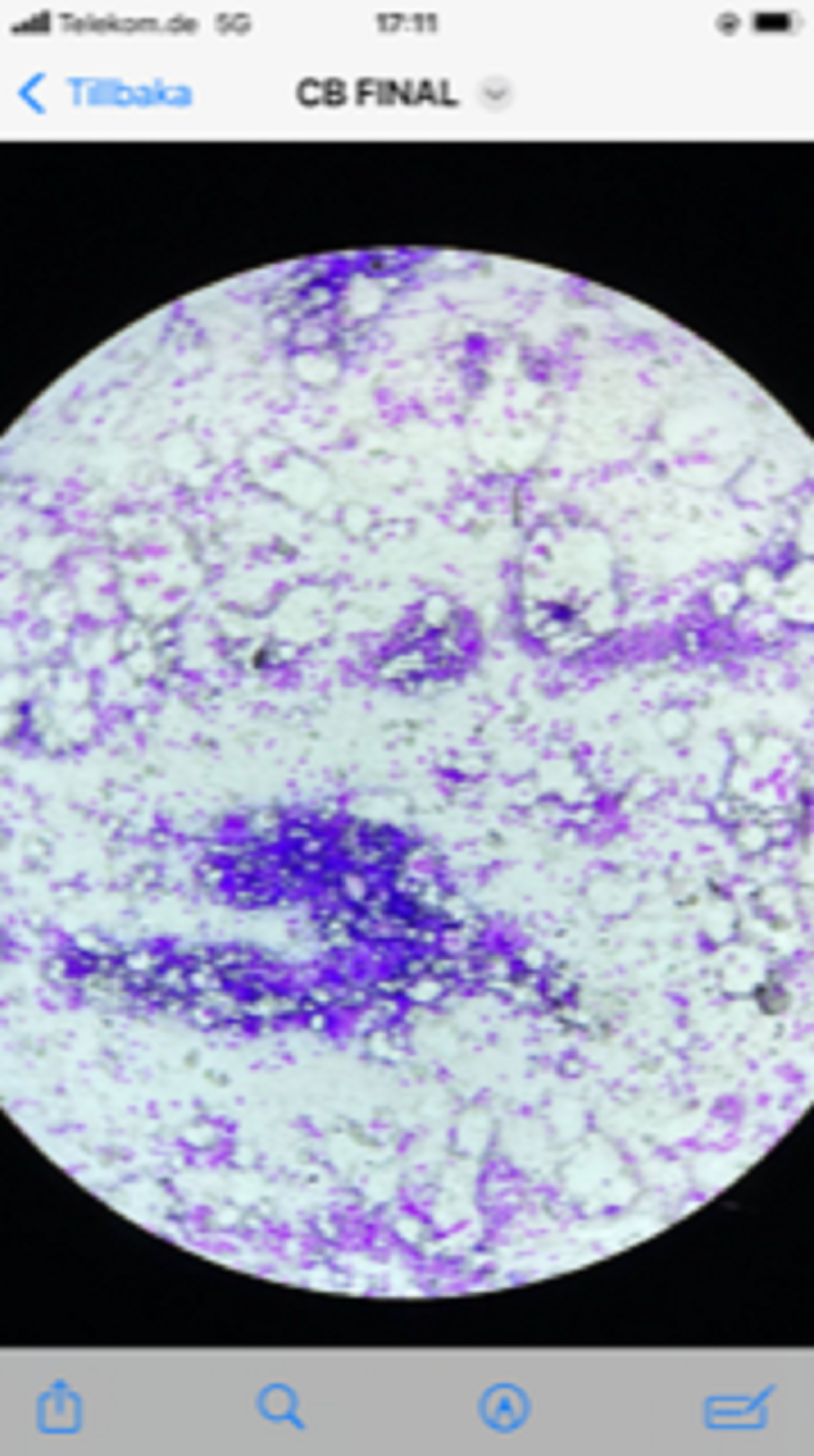
RER, Plasma cells, Bone marrow, MGG stain.
Reticulin fibres. Basement membrane. Nephrocytes. Renal tubules. Kidney. Silver impregnation
Golgi apparatus. Secretory neurons. Hypothalamus. Silver impregnation
Cilia and microvilli. Ependymal cells. Central canal. Spinal cord. HE stain
Collagen fibers. Skin. HE stain
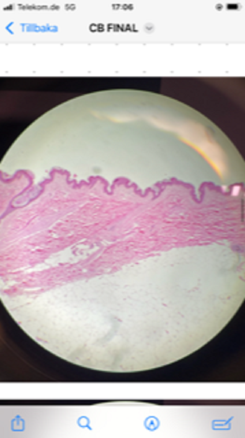
Collagen fibers. Skin. HE stain
Amitosis. Hepatocytes. Berg Bodies. Liver HE stain
Microvilli. Brush border. Nephrocytes. Kidney. HE stain
Stellate cells. Motor neurons. Spinal cord. H&E Stain
Stellate cells. Motor neurons. Spinal cord. H&E Stain
Basement membrane. Skin. PAS stain
Kinocilia. Epithelial cells. Trachea. HE stain
Elastic fibres. Artery cross section. Resorcin fuchsin stain
Reticulin fibres. Basement membrane. Nephrocytes. Renal tubules. Kidney. Silver impregnation
Mitosis. Bone marrow smear. MGG stain
Golgi apparatus. Secretory neurons. Hypothalamus. Silver impregnation
Stellate cells. Motor neurons. Spinal cord. H&E Stain
Basement membrane. Nephrocytes. Renal tubules. Kidney. PAS stain
Reticulin fibres. Basement membrane. Nephrocytes. Renal tubules. Kidney. Silver impregnation
Cilia and microvilli. Ependymal cells. Central canal. Spinal cord. HE stain
Kinocilia. Epithelial cells. Trachea. HE stain
Epididymis?
True
False
Testis?
True
False
{"name":"Cell biology quiz group 11", "url":"https://www.quiz-maker.com/QPREVIEW","txt":"Test your knowledge on cell biology with this comprehensive quiz designed for students and enthusiasts alike. With 40 thoughtfully crafted questions, you'll explore various aspects of cell structures, functions, and histological techniques.Whether you are preparing for exams or simply eager to learn more about the fascinating world of cells, this quiz is an excellent resource!40 QuestionsFocus on histology and cell functionMultichoice format for easy answering","img":"https:/images/course8.png"}
More Quizzes
Histo retake on entry cycle 1
221168
Histology test 2
10529
PSS Product Knowledge
15811
10 Types of People in the Office
1050
Free Direct vs Indirect Speech - Test Your Grammar
201032539
Hocus Pocus: Which Character Are You? Take It Now!
201023768
Macromolecular Solids
15821262
Free Hotel Product Knowledge
201025680
Challenge Yourself: Average Lifespan of a Red Blood Cell
201058211
Chicken Little: How Well Do You Know Abby Duck?
201029249
Tsunami Challenge: Ace 10 Key Tsunami Questions
201028026
What Should I Major In? Discover Your Best Degree
201023963