Unlock hundreds more features
Save your Quiz to the Dashboard
View and Export Results
Use AI to Create Quizzes and Analyse Results

Sign inSign in with Facebook
Sign inSign in with Google

Histology Identification Quiz: Sharpen Your Tissue ID Skills

Quick, free histology test to check your knowledge. Instant results.

Editorial: Review CompletedCreated By: Joy MorinUpdated Aug 28, 2025
Difficulty: Moderate
Questions: 20
Learning OutcomesStudy Material
Colorful paper art depicting cells and tissues for Histology Knowledge Assessment Quiz

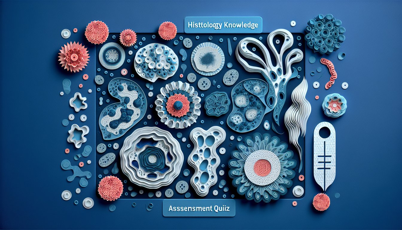
This histology quiz helps you practice tissue identification and common staining features with quick multiple-choice questions. See instant results and pinpoint topics to review. Keep learning with a histology practice quiz, focus on structures in a connective tissue quiz, or reinforce cell fundamentals in a cytology quiz.

Which epithelial type lines the alveoli of the lung, optimizing diffusion of gases?
Simple squamous epithelium
Transitional epithelium
Stratified squamous keratinized epithelium
Pseudostratified ciliated columnar epithelium
undefined
Which connective tissue fiber type provides tensile strength and is prominent in tendons?
Microfilaments
Elastic fibers
Collagen type I fibers
Reticular (collagen type III) fibers
undefined
Which cartilage type is found in the intervertebral discs and has abundant type I collagen?
Hyaline cartilage
Elastic cartilage
Fibrocartilage
Articular cartilage only
undefined
Which bone cell is primarily responsible for bone resorption?
Osteocyte
Chondrocyte
Osteoclast
Osteoblast
undefined
The structural unit of compact bone featuring concentric lamellae around a central canal is the
Lacuna
Volkmann canal
Osteon (Haversian system)
Canaliculus
undefined
Which muscle type has intercalated discs and centrally located nuclei?
Skeletal muscle
Myepithelial cells
Smooth muscle
Cardiac muscle
undefined
Which neuroglial cell myelinates axons in the central nervous system?
Oligodendrocyte
Microglia
Schwann cell
Astrocyte
undefined
Which epidermal layer contains keratohyalin granules and is prominent in thick skin?
Stratum spinosum
Stratum lucidum
Stratum granulosum
Stratum basale
undefined
In a sarcomere, which band shortens during contraction?
I band
Z line
M line
A band
undefined
Which structure surrounds individual nerve fibers in a peripheral nerve?
Basement membrane
Endoneurium
Perineurium
Epineurium
undefined
In the spleen, lymphoid nodules around central arterioles are part of the
Trabeculae
Marginal sinus only
Red pulp
White pulp
undefined
Which cells form the visceral layer of Bowman capsule with foot processes?
Macula densa cells
Podocytes
Juxtaglomerular cells
Mesangial cells
undefined
The juxtaglomerular apparatus includes juxtaglomerular cells and which specialized epithelial patch?
Brush border
Podocyte diaphragm
Macula densa
Principal cells
undefined
In the classic hepatic lobule concept, blood flows toward which central structure?
Portal vein
Bile duct
Central vein
Hepatic artery
undefined
Which stage of erythropoiesis first shows loss of the nucleus?
Basophilic erythroblast
Reticulocyte
Proerythroblast
Orthochromatophilic erythroblast
undefined
Which feature distinguishes the jejunum from duodenum and ileum histologically?
Absence of villi
Tall plicae circulares with long villi, few Brunner glands and Peyer patches
Numerous Peyer patches in submucosa/lamina propria
Abundant Brunner glands in submucosa
undefined
Which anterior pituitary cell type predominantly secretes growth hormone and stains acidophilic?
Thyrotroph
Somatotroph
Corticotroph
Gonadotroph
undefined
Placental villi at term are best described as
Cartilage-containing villi
Highly branched villi with thin syncytiotrophoblast and numerous fetal capillaries
Primary villi with cytotrophoblast core only
Villi lacking trophoblast layers
undefined
Which fiber type is best visualized with silver stains and forms a supporting network in lymphoid organs?
Microtubules
Type I collagen bundles
Reticular fibers (type III collagen)
Elastic fibers
undefined
Which component of the basement membrane contains type IV collagen and laminin and is produced by epithelial cells?
Basal lamina
External lamina of muscle cells
Elastic lamina
Reticular lamina
undefined
0

Learning Outcomes

  1. Analyse cell and tissue microstructures under microscopy
  2. Identify key histological features across different tissue types
  3. Interpret staining patterns for diagnostic accuracy
  4. Apply classification criteria to epithelial and connective tissues
  5. Evaluate histological slides with critical reasoning

Cheat Sheet

  1. Primary tissue types - Your body is built from four superstar tissue types: epithelial, connective, muscle, and nervous. Each one brings its own special structure and function, from forming protective barriers to sending electric signals at lightning speed. Ready to meet these cellular heroes?
  2. Hematoxylin & Eosin staining - Discover how H&E staining paints a vivid picture of nuclei and cytoplasm, making it easier to spot cells and understand their layout. This classic technique is your microscope's best friend when it comes to telling different tissues apart.
  3. Key histological features - Train your eyes to notice unique patterns like layered epithelial cells or the web of fibers in connective tissue. Identifying these signatures is crucial for distinguishing one tissue from another.
  4. Microscope practice - Grab your slides and get hands-on: the more you analyze normal tissue under the scope, the quicker you'll spot anything unusual. Consistent practice turns those tiny shapes into big "aha!" moments.
  5. Tissue preparation process - From fixation and embedding to slicing thin sections and staining, each step matters for creating clear, informative slides. Understanding the workflow helps you appreciate why samples look the way they do under the lens.
  6. Epithelial classification - Get to know squamous, cuboidal, and columnar cell shapes plus the difference between simple and stratified layers. Mastering these categories is like having a secret code for identifying linings and barriers in the body.
  7. Connective tissue characteristics - Dive into the world of collagen, elastic, and reticular fibers, and meet the cells that keep everything in place - fibroblasts, adipocytes, and more. Spotting these elements is key to understanding support structures throughout the body.
  8. Muscle tissue organization - Learn to tell skeletal, cardiac, and smooth muscle apart by their striations, branch patterns, and control types. This knowledge helps you link microscopic structure to powerful contractions and rhythmic beats.
  9. Nervous tissue components - Explore the intricate architecture of neurons, with their long axons and branching dendrites, alongside glial cells that support and protect. Recognizing these details reveals how signals travel through your entire nervous system.
  10. Critical reasoning with slides - Don't just look - think! Compare your observations to normal structures, consider physiological roles, and predict what pathologies might cause deviations. This analytical approach sharpens both your microscope skills and diagnostic mindset.
Powered by: Quiz Maker