Unlock hundreds more features
Save your Quiz to the Dashboard
View and Export Results
Use AI to Create Quizzes and Analyse Results

Sign inSign in with Facebook
Sign inSign in with Google

Skull Labeling Quiz: Identify Cranial Bones and Landmarks

Quick, free cranium labeling quiz. Instant feedback to track progress.

Editorial: Review CompletedCreated By: Danaya ChittratanawatUpdated Aug 26, 2025
Difficulty: Moderate
Grade: Grade 10
Study OutcomesCheat Sheet
Paper art depicting trivia quiz, Skull Label Challenge, for high school biology students.

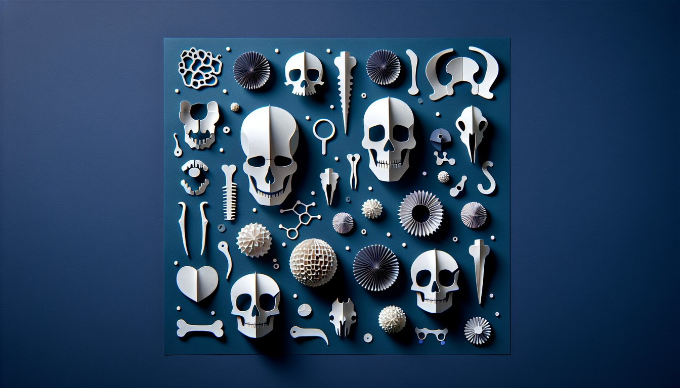
This skull labeling quiz helps you practice identifying the bones of the cranium and key landmarks, building speed and accuracy for class or exams. After you finish, keep learning with a focused cranial bones quiz, explore regional detail in the skull and facial anatomy quiz, or round out your axial anatomy with the vertebrae labeling quiz.

Which bone forms the forehead and the roof of the orbits?
Frontal bone
Maxilla
Zygomatic bone
Parietal bone
undefined
The parietal bones form a single unpaired bone.
False
True
undefined
The temporal bone houses structures of the inner ear.
True
False
undefined
The maxilla forms the lower jaw.
False
True
undefined
The mandible is the only movable bone of the skull (excluding the auditory ossicles).
True
False
undefined
Which structures of the occipital bone articulate with the atlas (C1) to allow nodding?
External occipital protuberance
Occipital condyles
Foramen magnum
Nuchal lines
undefined
The nasal bones form the lower margin of the orbit.
False
True
undefined
Which tiny facial bone lies in the medial wall of the orbit and contains the nasolacrimal groove?
Nasal bone
Ethmoid bone
Palatine bone
Lacrimal bone
undefined
The vomer forms the inferior portion of the nasal septum.
True
False
undefined
The sella turcica of the sphenoid bone houses the pineal gland.
False
True
undefined
The cribriform plate of the ethmoid bone transmits olfactory nerve fibers from the nasal cavity to the cranial cavity.
False
True
undefined
Which anterior projection of the mandible serves as the insertion for the temporalis muscle?
Mandibular notch
Condylar process
Mental spine
Coronoid process
undefined
Which bone contributes to the posterior wall of the orbit and contains the optic canal?
Maxilla
Zygomatic bone
Nasal bone
Sphenoid bone
undefined
Which paired, downward-projecting processes arise from the sphenoid bone and provide muscle attachment near the nasal cavity?
Pterygoid processes
Styloid processes
Zygomatic processes
Mastoid processes
undefined
Which vertical midline projection of the ethmoid bone provides attachment for the falx cerebri?
Cribriform plate
Crista ampullaris
Crista galli
Perpendicular plate
undefined
Which large foramen, located between the temporal and occipital bones, transmits the internal jugular vein and cranial nerves IX, X, and XI?
Foramen ovale
Foramen lacerum
Foramen rotundum
Jugular foramen
undefined
Which fissure in the orbit transmits cranial nerves III, IV, V1, and VI?
Optic canal
Inferior orbital fissure
Foramen ovale
Superior orbital fissure
undefined
Which structures form the zygomatic arch?
Zygomatic process of the mandible and temporal process of the maxilla
Temporal process of the maxilla and zygomatic process of the frontal bone
Zygomatic process of the maxilla and frontal process of the zygomatic bone
Zygomatic process of the temporal bone and temporal process of the zygomatic bone
undefined
Which foramen in the sphenoid bone transmits the mandibular division of the trigeminal nerve (V3)?
Foramen ovale
Foramen spinosum
Foramen rotundum
Foramen lacerum
undefined
Which weak point on the lateral skull overlies the middle meningeal artery and is clinically significant in epidural hematomas?
Asterion
Glabella
Inion
Pterion
undefined
0

Study Outcomes

  1. Identify key anatomical features of the skull through label matching.
  2. Recall the names and functions of important cranial structures.
  3. Analyze the spatial relationships between different skull parts.
  4. Apply learned knowledge to correctly label skull diagrams during tests.

Label Skull Review Cheat Sheet

  1. Skull Composition - The human skull is like a 22-piece puzzle: 8 cranial bones form the protective braincase while 14 facial bones create your unique facial features. Getting comfy with this bone count is your first power move in skull anatomy.
  2. Frontal Bone - Think of the frontal bone as your forehead's VIP; it shapes the brow ridge and the upper eye sockets. Spotting this bone helps you map out its neighboring structures in no time.
  3. Parietal Bones - These two mirror‑image bones sit on each side of your skull, meeting at the sagittal suture like long‑lost twins. Learning this seam is crucial for understanding skull symmetry and growth.
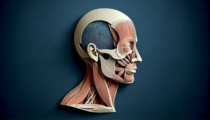
  4. Temporal Bones - Your temporal bones house delicate ear structures and form the skull's sturdy sidewalls. Pinpointing these bones unlocks the secrets of hearing and balance.
  5. Occipital Bone - This powerhouse bone makes up the back and base of the skull, complete with the foramen magnum where the spinal cord waves hello to the brain. Recognizing it is key for understanding brainstem connections.
  6. Sphenoid Bone - Hidden at the skull's base, the sphenoid bone plays matchmaker by connecting to almost every other cranial bone and cradles the pituitary gland in the sella turcica. Its central spot makes it a superstar in skull architecture.
  7. Ethmoid Bone - Sneaky and lightweight, the ethmoid bone contributes to both your nasal cavity and eye sockets. Grasping its location helps you tie together respiratory and visual pathways.
  8. Coronal Suture - This zigzag line splits the forehead's frontal bone from the twin parietal bones, much like a crown's edge. Spotting it gives you major insight into skull segmentation and growth patterns.
  9. Squamosal Suture - Running along the side of your skull, the squamosal suture joins the temporal and parietal bones. It's a great landmark for understanding the skull's lateral architecture.
  10. Lambdoid Suture - At the back of the skull, this suture weaves between the occipital and parietal bones, resembling an upside‑down "V." Learning it ties together your knowledge of the skull's rear structure.
Powered by: Quiz Maker