Unlock hundreds more features
Save your Quiz to the Dashboard
View and Export Results
Use AI to Create Quizzes and Analyse Results

Sign inSign in with Facebook
Sign inSign in with Google

Abdominal Muscles Quiz: Test Your Core Anatomy

Quick abdominal anatomy quiz to test your core knowledge. Instant results.

Editorial: Review CompletedCreated By: Tanel VoorelUpdated Aug 24, 2025
Difficulty: Moderate
2-5mins
Learning OutcomesCheat Sheet
Paper art abdominal muscles layered on sky blue background with quiz title and labels hinting core anatomy challenge

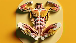
This abdominal muscles quiz helps you check your knowledge of the rectus abdominis, obliques, and other core muscles, including actions and locations. Get instant results and see what to review next. Want more practice? Try an abdomen labeling activity, explore an abdominal anatomy quiz, or round it out with a back muscles quiz.

Which muscle is the most superficial of the anterolateral abdominal wall?
Internal oblique
External oblique
Transversus abdominis
Rectus abdominis
The external oblique is the most superficial muscle of the anterolateral abdominal wall, with fibers running inferomedially toward the linea alba. It overlies the internal oblique and transversus abdominis muscles. Its broad aponeurosis contributes to the formation of the anterior rectus sheath. .
The "six-pack" appearance of the abdomen is created by the tendinous intersections of which muscle?
Rectus abdominis
Transversus abdominis
Internal oblique
External oblique
The tendinous intersections segment the rectus abdominis into distinct sections, producing the characteristic "six-pack" appearance. These tendinous bands anchor the muscle to the anterior abdominal wall. Only the rectus abdominis has this segmented structure. .
Which structure runs vertically down the midline of the anterior abdominal wall?
Arcuate line
Inguinal ligament
Linea alba
Linea semilunaris
The linea alba is a fibrous structure running vertically along the midline of the anterior abdominal wall, formed by the fusion of the aponeuroses of the abdominal muscles. It extends from the xiphoid process to the pubic symphysis. It serves as an important landmark in abdominal surgeries. .
Which muscle's fibers run horizontally (transversely) across the abdomen?
Internal oblique
External oblique
Rectus abdominis
Transversus abdominis
The transversus abdominis has fibers that run horizontally around the abdominal wall, acting like a corset to compress abdominal contents. This orientation distinguishes it from the oblique and rectus muscles. It is the deepest of the anterolateral abdominal muscles. .
The inguinal ligament is the thickened lower border of which muscle's aponeurosis?
External oblique
Internal oblique
Transversus abdominis
Rectus abdominis
The inguinal ligament is formed by the rolled-under inferior border of the external oblique aponeurosis, extending from the anterior superior iliac spine to the pubic tubercle. It forms the floor of the inguinal canal. .
Which nerve root predominantly innervates the skin around the umbilicus?
T10
T12
L1
T9
The T10 spinal nerve root supplies the dermatome that includes the skin around the umbilicus. This landmark is commonly used in clinical examinations to assess nerve function. .
The linea semilunaris marks the lateral border of which muscle?
Transversus abdominis
External oblique
Rectus abdominis
Internal oblique
The linea semilunaris is a curved tendinous line marking the lateral border of the rectus abdominis muscle on the anterior abdominal wall. It results from the aponeurotic junction of the abdominal muscles. .
Which abdominal muscle's primary function is to compress the abdominal contents?
Transversus abdominis
Internal oblique
External oblique
Rectus abdominis
The transversus abdominis acts like a corset to compress abdominal viscera, increasing intra-abdominal pressure and stabilizing the trunk. It is the deepest of the abdominal muscles with horizontal fibers. .
The external oblique muscle originates from which ribs?
3rd to 10th ribs
5th to 12th ribs
7th to 12th ribs
1st to 8th ribs
The external oblique originates from the external surfaces of the 5th to 12th ribs. Its fibers run inferomedially toward the linea alba. .
Contraction of the external oblique on one side results in what trunk movement?
Lateral flexion
Contralateral rotation
Extension
Ipsilateral rotation
Unilateral contraction of the external oblique causes contralateral rotation of the trunk, pulling the thorax toward the opposite side. The internal oblique, in contrast, produces ipsilateral rotation. .
At what level does the arcuate line of the rectus sheath typically lie?
Just below the costal margin
Halfway between the umbilicus and pubic symphysis
At the xiphoid process
Level of the iliac crest
The arcuate line is typically located about halfway between the umbilicus and the pubic symphysis. Below this line, all aponeurotic layers pass anterior to the rectus abdominis. .
Which muscle inserts onto the pubic crest and pectineal line via the conjoint tendon?
Rectus abdominis
Transversus abdominis
Internal oblique
External oblique
The conjoint tendon is formed by the fusion of the internal oblique and transversus abdominis aponeuroses. It inserts onto the pubic crest and pectineal line, reinforcing the posterior wall of the inguinal canal. .
The transversalis fascia is located between which two structures?
Internal oblique and transversus abdominis
External oblique and internal oblique
Rectus abdominis and transversus abdominis
Transversus abdominis and parietal peritoneum
The transversalis fascia lies deep to the transversus abdominis muscle and superficial to the parietal peritoneum, forming part of the posterior abdominal wall. It provides a layer of connective tissue support. .
The deep inguinal ring is an opening in which layer of the abdominal wall?
Transversalis fascia
Internal oblique muscle
External oblique aponeurosis
Transversus abdominis muscle
The deep inguinal ring is a defect in the transversalis fascia located superior to the inguinal ligament. It marks the entrance to the inguinal canal. .
Which artery supplies blood to the lower part of the rectus abdominis muscle?
Inferior epigastric artery
Deep circumflex iliac artery
Lateral thoracic artery
Superior epigastric artery
The inferior epigastric artery, branching from the external iliac artery, ascends on the posterior surface of the rectus abdominis to supply its lower portion. It anastomoses with the superior epigastric artery above. .
The thoracoabdominal nerves that innervate the anterolateral abdominal muscles are continuations of which intercostal nerves?
T12 to L1
T1 to T5
T7 to T11
L2 to L4
The thoracoabdominal nerves are the anterior rami of the lower five intercostal nerves (T7 - T11). They provide motor and sensory innervation to the anterolateral abdominal wall. .
Above the arcuate line, which aponeuroses form the posterior wall of the rectus sheath?
Transversus abdominis and external oblique
External oblique and internal oblique
Only transversus abdominis
Internal oblique and transversus abdominis
Above the arcuate line, the posterior layer of the rectus sheath is formed by the aponeuroses of the internal oblique (which splits around rectus) and the transversus abdominis. The external oblique aponeurosis contributes only to the anterior sheath. .
The upper portion of the rectus abdominis is primarily innervated by which nerves?
L1 to L2
T1 to T6
T10 to T12
T7 to T9
The upper fibers of the rectus abdominis muscle receive segmental innervation from the thoracoabdominal nerves T7 to T9. The lower fibers are innervated by T10 to T12. .
A Spigelian hernia occurs through a defect in which anatomical structure?
Linea alba near the xiphoid process
Linea semilunaris at the level of the arcuate line
Inguinal canal floor
Femoral canal
A Spigelian hernia occurs through a defect in the linea semilunaris, typically at or below the arcuate line where the posterior rectus sheath is absent. This allows abdominal contents to protrude. .
The conjoint tendon contributes to the posterior wall of which region of the inguinal canal?
Supravesical fossa
Lateral inguinal (deep ring) region
Medial inguinal (Hesselbach's) triangle
Femoral canal
The conjoint tendon, formed by fused aponeuroses of the internal oblique and transversus abdominis, reinforces the medial posterior wall of the inguinal canal, particularly Hesselbach's triangle. .
Which ligament attaches to the pectineal line of the pubis and is formed by a reflection of the inguinal ligament?
Reflected inguinal ligament
Inguinal ligament
Pectineal (Cooper's) ligament
Lacunar (Gimbernat's) ligament
The pectineal or Cooper's ligament is formed by the lateral extension of the lacunar ligament along the pectineal line of the pubis. It provides reinforcement to the femoral canal. .
The neurovascular plane between the internal oblique and transversus abdominis muscles contains which nerve?
Subcostal nerve
Iliohypogastric nerve
Ilioinguinal nerve
Inferior epigastric nerve
The iliohypogastric nerve travels in the plane between the internal oblique and transversus abdominis muscles, providing sensory innervation to the skin of the lower abdomen. .
The small pyramidalis muscle, when present, tenses which structure?
Transversalis fascia
Conjoint tendon
Inguinal ligament
Linea alba
The pyramidalis muscle tenses the linea alba by pulling it downward when it contracts. It lies anterior to the lower part of the rectus abdominis. .
Which layer of the thoracolumbar fascia lies anterior to the quadratus lumborum muscle?
Middle layer
Transversalis fascia
Posterior layer
Anterior layer
The anterior layer of the thoracolumbar fascia lies in front of the quadratus lumborum muscle, separating it from the posterior abdominal wall. The middle and posterior layers have different attachments. .
During a transversus abdominis plane (TAP) block, local anesthetic is deposited between which two muscle layers?
Rectus abdominis and external oblique
Internal oblique and transversus abdominis
External and internal oblique
Transversus abdominis and transversalis fascia
In a TAP block, local anesthetic is injected into the plane between the internal oblique and transversus abdominis muscles to anesthetize the thoracoabdominal nerves. This technique provides effective analgesia for abdominal surgery. .
0
{"name":"Which muscle is the most superficial of the anterolateral abdominal wall?", "url":"https://www.quiz-maker.com/QPREVIEW","txt":"Which muscle is the most superficial of the anterolateral abdominal wall?, The \"six-pack\" appearance of the abdomen is created by the tendinous intersections of which muscle?, Which structure runs vertically down the midline of the anterior abdominal wall?","img":"https://www.quiz-maker.com/3012/images/ogquiz.png"}

Study Outcomes

  1. Identify Major Abdominal Muscles -

    Recognize and name key muscles in the abdominal wall, including the rectus abdominis, external and internal obliques, and transverse abdominis.

  2. Describe Muscle Functions -

    Explain the primary actions of each abdominal muscle, such as trunk flexion, rotation, and core stabilization.

  3. Differentiate Anatomical Layers -

    Distinguish between superficial and deep layers of the muscles of the abdomen and understand their spatial relationships.

  4. Apply Core Anatomy Knowledge -

    Use your understanding of trunk anatomy to answer quiz questions accurately and reinforce your core muscle quiz skills.

  5. Relate Anatomy to Function -

    Connect abdominal muscle structure to practical applications like posture support and movement efficiency.

  6. Evaluate Knowledge Gaps -

    Assess your quiz performance to identify areas for further study in core and trunk anatomy.

Cheat Sheet

  1. Rectus Abdominis Structure -

    The rectus abdominis runs vertically from the pubic crest to the xiphoid process and is segmented by three tendinous intersections, allowing prominent "six-pack" definition. A helpful mnemonic is "Ripples on the Rib-to-Pubis Path" to recall its attachments, as noted in Gray's Anatomy. Mastering this layout will boost confidence when tackling the abdominal muscles quiz on trunk flexion.

  2. Oblique Muscle Fiber Directions -

    External oblique fibers run inferomedially (think "hands in pockets") while internal oblique fibers run superomedially (reverse "hands on heart"), enabling rotation and lateral flexion. This hand-in-pocket mnemonic is endorsed by the American Council on Exercise for core muscle quiz prep. Recognizing these cross-hatch patterns makes spine rotation questions in a trunk anatomy quiz a breeze.

  3. Transversus Abdominis Activation -

    The deepest abdominal wall muscle has horizontal fibers that compress the abdominal cavity and stabilize the spine; remember "TV for TransVerse" to visualize its belt-like wrap. According to studies from Stanford's biomechanical lab, engaging the transversus abdominis with the "draw-in" maneuver improves core stability. Practicing this will reinforce your understanding in an abdominal wall muscles quiz.

  4. Linea Alba and Sheath Composition -

    The linea alba is a fibrous midline seam formed by the fusion of the rectus sheath aponeuroses, with distinct anterior and posterior layers above the arcuate line. Use the rule "EPA Above, EAAnterior Below" (External, Internal, Transverse Aponeuroses above; only External and portion of Internal below) from standard anatomy texts. This clarity is key for acing questions on the muscles of the abdomen quiz regarding load transmission.

  5. Innervation and Blood Supply -

    Segmental innervation comes from T7 - T12 intercostal and L1 iliohypogastric/ilioinguinal nerves, while superior and inferior epigastric vessels provide key blood flow. A handy phrase is "7 up to L1" for nerve levels and "SEI for Superior, EII for Inferior Epigastrics" as taught by the National Library of Medicine. Cementing these details ensures you'll sail through the core muscle quiz vascular questions!

Powered by: Quiz Maker