Unlock hundreds more features
Save your Quiz to the Dashboard
View and Export Results
Use AI to Create Quizzes and Analyse Results

Sign inSign in with Facebook
Sign inSign in with Google

Abdominal Muscles Quiz: Can You Identify Them All?

Think you can ace this abdominal wall muscle quiz? Label every muscle now!

Difficulty: Moderate
2-5mins
Learning OutcomesCheat Sheet
Paper art illustration for abdominal muscles quiz on golden yellow background

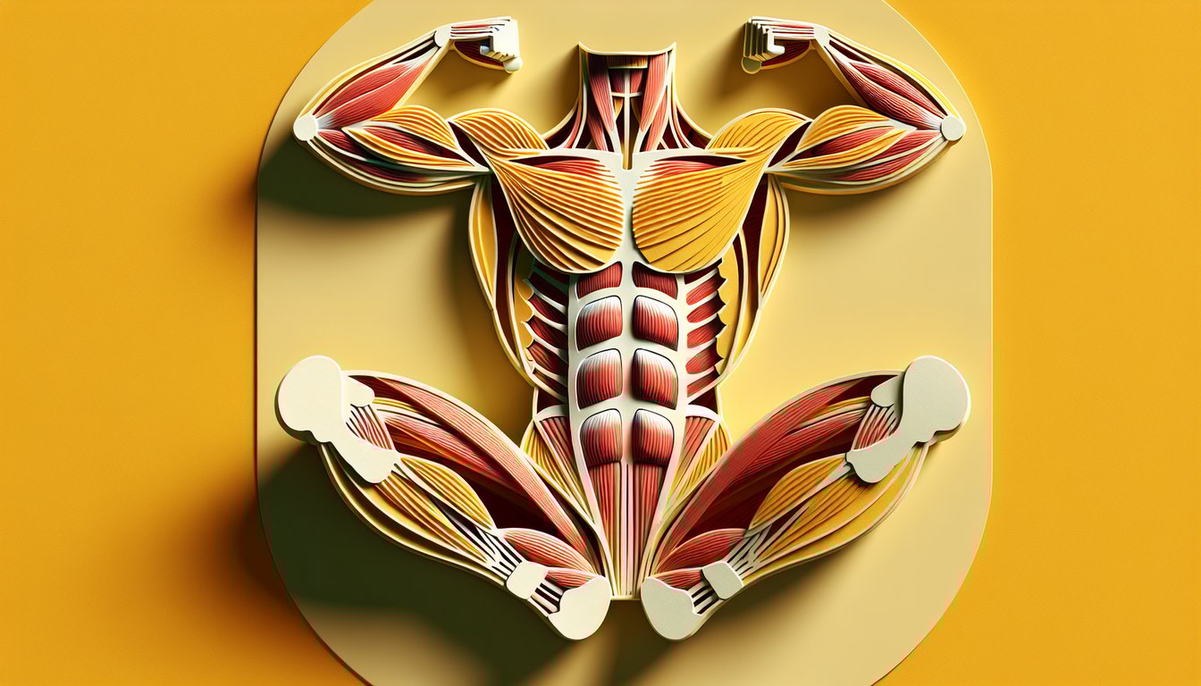
This abdominal muscles quiz helps you label every muscle of the abdominal wall on clear diagrams. Use this quiz and practice labeling each muscle to spot gaps before class or an exam, with instant feedback to build confidence. You'll cover rectus abdominis, obliques, and transversus abdominis.

Which muscle is the most superficial layer of the anterior abdominal wall?
Transversus abdominis
External oblique
Internal oblique
Rectus abdominis
The external oblique forms the outermost layer of the anterior abdominal wall and has fibers running inferomedially. It aids in trunk rotation and compression of abdominal contents. Beneath it lies the internal oblique, followed by the transversus abdominis.
Which abdominal muscle originates from the outer surfaces of ribs 5 - 12?
Internal oblique
Quadratus lumborum
External oblique
Transversus abdominis
The external oblique muscle arises from the outer surfaces of ribs five through twelve. Its broad aponeurosis contributes to the linea alba and inguinal ligament. This orientation allows it to assist in trunk flexion and rotation.
Which abdominal muscle's fibers run transversely across the abdomen?
Rectus abdominis
External oblique
Internal oblique
Transversus abdominis
The transversus abdominis has fibers that run horizontally (transversely) around the abdomen, acting like a corset. It is the deepest of the flat abdominal muscles and plays a key role in core stability and increasing intra?abdominal pressure. Its aponeurosis contributes to the posterior layer of the rectus sheath above the arcuate line.
Which structure is formed by the fusion of the aponeuroses of the abdominal muscles at the midline?
Linea semilunaris
Inguinal ligament
Arcuate line
Linea alba
The linea alba is a fibrous raphe formed by the interlacing aponeuroses of the external oblique, internal oblique, and transversus abdominis muscles. It runs vertically from the xiphoid process to the pubic symphysis. It is a common surgical entry point due to its relative avascularity.
Which muscle is commonly referred to as the 'six-pack' due to its segmented appearance?
External oblique
Pyramidalis
Transversus abdominis
Rectus abdominis
The rectus abdominis is segmented by tendinous inscriptions and gives the appearance of a 'six-pack'. It runs vertically on each side of the linea alba. Its primary action is trunk flexion and stabilization during movement.
Which abdominal muscle lies immediately deep to the external oblique and has fibers running superomedially?
Internal oblique
Rectus abdominis
Transversus abdominis
External oblique
The internal oblique lies directly beneath the external oblique, with fibers running superomedially toward the linea alba. It assists in trunk rotation and lateral flexion. Its aponeurosis contributes to both anterior and posterior layers of the rectus sheath above the arcuate line.
As part of its actions, which muscle primarily compresses the abdominal contents?
Transversus abdominis
External oblique
Internal oblique
Rectus abdominis
The transversus abdominis wraps around the abdomen and, when it contracts, acts like a belt to compress the abdominal contents. This action increases intra?abdominal pressure, important for forced expiration, defecation, and childbirth. It also stabilizes the lumbar spine.
Where is the linea alba located?
Lateral border of the rectus abdominis
Midline between the two rectus abdominis muscles
Between external and internal oblique muscles
At the superficial inguinal ring
The linea alba is a midline fibrous raphe formed by the fusion of the aponeuroses of the flat abdominal muscles. It extends vertically from the xiphoid process to the pubic symphysis. It separates the left and right rectus abdominis muscles.
Which nerves provide motor innervation to the external oblique muscle?
Obturator nerve (L2 - L4)
Iliohypogastric nerve (L1)
Thoracoabdominal nerves (T7 - T11)
Femoral nerve (L2 - L4)
The external oblique is innervated by the thoracoabdominal nerves, which are continuations of the intercostal nerves T7 through T11. These nerves travel between the internal oblique and transversus abdominis before reaching the external oblique. The iliohypogastric (L1) nerve supplies part of the internal oblique and transversus abdominis, not the external layer.
At which vertebral level does the transumbilical plane lie?
T10 - T11
L1 - L2
L5 - S1
L3 - L4
The transumbilical plane is a horizontal anatomical plane that passes through the umbilicus at the level of vertebral bodies L3 to L4. It divides the abdomen into quadrants for clinical reference. It is frequently used as a landmark in abdominal surgery and examination.
Which artery supplies the superior portion of the rectus abdominis muscle?
Deep circumflex iliac artery
Inferior epigastric artery
Superior epigastric artery
Superficial epigastric artery
The superior epigastric artery is a continuation of the internal thoracic artery and enters the rectus sheath to supply the upper part of the rectus abdominis. It anastomoses with the inferior epigastric artery around the umbilical region. This dual supply helps maintain blood flow if one artery is compromised.
Which artery anastomoses with the superior epigastric artery within the rectus sheath?
Deep circumflex iliac artery
Superficial epigastric artery
Inferior epigastric artery
Superior epigastric artery
Within the rectus sheath, the inferior epigastric artery ascends to meet the superior epigastric artery, forming an important anastomosis. The inferior epigastric originates from the external iliac artery. This connection provides collateral circulation to the anterior abdominal wall.
Through which opening of the abdominal wall does the spermatic cord pass?
Deep inguinal ring
Superficial inguinal ring
Femoral canal
Obturator canal
The deep inguinal ring is an opening in the transversalis fascia lateral to the inferior epigastric vessels and is the entrance to the inguinal canal. The spermatic cord passes through this ring from the abdominal cavity. It then exits via the superficial inguinal ring.
Which small triangular muscle, when present, tenses the linea alba?
Quadratus lumborum
Rectus abdominis
Transversus abdominis
Pyramidalis
The pyramidalis is a small, triangular muscle located anterior to the lower part of the rectus abdominis. It tenses the linea alba when it contracts. The muscle is absent in approximately 20% of individuals.
What tendon is formed by the fusion of the internal oblique and transversus abdominis aponeuroses?
Inguinal ligament
Linea alba
Conjoint tendon
Rectus sheath
The conjoint tendon (falx inguinalis) is formed by the fusion of the lower fibers of the internal oblique and transversus abdominis aponeuroses. It reinforces the posterior wall of the inguinal canal near the superficial inguinal ring. It is clinically significant in understanding direct inguinal hernias.
What defines the arcuate line in the abdominal wall?
Lower limit of the posterior layer of the rectus sheath
Attachment of the inguinal ligament
Upper limit of the linea alba
Lateral border of the rectus abdominis
The arcuate line marks the lower border of the posterior layer of the rectus sheath. Below this line, all aponeuroses of the flat abdominal muscles pass anterior to the rectus abdominis, leaving only the transversalis fascia posteriorly. It is clinically relevant for surgical approaches.
Which layer of the rectus sheath is absent below the arcuate line?
Posterior layer
Anterior layer
Rectus abdominis muscle
Transversalis fascia
Below the arcuate line, the posterior layer of the rectus sheath is absent as all three aponeuroses pass anterior to the rectus abdominis. The transversalis fascia lies directly posterior to the rectus muscle in this region. This anatomical change has implications for hernia risk and surgical planning.
What structure forms the posterior boundary of the inguinal canal?
Internal oblique muscle
External oblique aponeurosis
Inguinal ligament
Transversalis fascia
The transversalis fascia constitutes the posterior wall of the inguinal canal throughout its length. It is reinforced medially by the conjoint tendon near the superficial inguinal ring. The external oblique aponeurosis forms the anterior wall instead.
Which nerve runs between the internal oblique and transversus abdominis muscles?
Femoral nerve
Obturator nerve
Iliohypogastric nerve
Subcostal nerve
The iliohypogastric nerve emerges from L1 and travels between the internal oblique and transversus abdominis muscles. It provides sensory innervation to the skin above the pubis and motor fibers to the lower abdominal muscles. The subcostal nerve (T12) runs just above it.
What is the origin of the pyramidalis muscle?
Iliac crest
Pubic crest
Pubic tubercle
Xiphoid process
The pyramidalis muscle originates from the front of the pubic crest and the pubic symphysis. It inserts into the linea alba and tenses it upon contraction. This small muscle is absent in a significant minority of individuals.
Which muscle plays the primary role in compressing the abdominal cavity to increase intra-abdominal pressure?
External oblique
Rectus abdominis
Internal oblique
Transversus abdominis
The transversus abdominis, as the deepest layer of the abdominal wall, acts like a natural corset to compress the abdominal contents and increase intra?abdominal pressure. This is crucial during forced expiration, coughing, and Valsalva maneuvers. Its tonic activation also stabilizes the lumbar spine.
Which dermatome level corresponds to sensation at the umbilicus?
T8
L1
T10
T12
The umbilicus is innervated by the T10 dermatome, which is a key surface landmark in physical examination. Pain from appendicitis initially presents in this periumbilical region. Understanding dermatomal levels aids in diagnosing visceral and referred pain.
Which superficial fascia layer is continuous with the Dartos fascia of the scrotum?
Camper's fascia
Endoabdominal fascia
Transversalis fascia
Scarpa's fascia
Scarpa's fascia is the deep membranous layer of superficial fascia in the lower anterior abdominal wall. It is continuous with Dartos fascia of the scrotum and Colles fascia of the perineum. Camper's fascia is the fatty superficial layer instead.
Which aponeurosis contributes to the anterior wall of the inguinal canal?
Internal oblique aponeurosis
External oblique aponeurosis
Rectus sheath aponeurosis
Transversus abdominis aponeurosis
The anterior wall of the inguinal canal is formed mainly by the aponeurosis of the external oblique muscle. The internal oblique fibers contribute to the roof, while the conjoint tendon reinforces the posterior wall. Understanding these layers is critical in hernia repair.
Which spinal nerve roots form the ilioinguinal nerve?
T10 - T11
L2
L1
T12 - L1
The ilioinguinal nerve arises from the L1 spinal nerve root after it exits the spinal canal. It travels through the inguinal canal to supply sensation to the upper medial thigh and parts of the genitalia. The subcostal nerve (T12) runs separately but often joins closely.
Which fascial layer must be cut to enter the preperitoneal space during a laparoscopic TEP inguinal hernia repair?
Transversalis fascia
External oblique aponeurosis
Internal oblique muscle
Scarpa's fascia
In a Totally Extraperitoneal (TEP) repair, the surgeon dissects between the peritoneum and the abdominal wall by incising the transversalis fascia. This creates the preperitoneal space for mesh placement without entering the peritoneal cavity. The external oblique aponeurosis is not breached in the TEP approach.
In a Spigelian hernia, protrusion occurs through which anatomical landmark?
Posterior rectus sheath
Superficial inguinal ring
Linea alba
Semilunar line at the level of the arcuate line
A Spigelian hernia occurs along the semilunar (Spigelian) line, typically just below or at the level of the arcuate line where the posterior rectus sheath is deficient. It protrudes through a defect in the transversus aponeurosis. These hernias are often interparietal and can be difficult to detect clinically.
Within the posterior rectus sheath above the arcuate line, where is the inferior epigastric artery located relative to the rectus abdominis muscle?
Outside the sheath in the extraperitoneal fat
Deep to the muscle on the posterior sheath
Superficial to the muscle on the anterior sheath
Lateral to the muscle between oblique muscles
Above the arcuate line, the inferior epigastric artery travels deep to the rectus abdominis muscle within the posterior layer of its sheath. It then ascends to anastomose with the superior epigastric artery. Below the arcuate line it lies between the muscle and transversalis fascia.
0
{"name":"Which muscle is the most superficial layer of the anterior abdominal wall?", "url":"https://www.quiz-maker.com/QPREVIEW","txt":"Which muscle is the most superficial layer of the anterior abdominal wall?, Which abdominal muscle originates from the outer surfaces of ribs 5 - 12?, Which abdominal muscle's fibers run transversely across the abdomen?","img":"https://www.quiz-maker.com/3012/images/ogquiz.png"}

Study Outcomes

  1. Identify Abdominal Wall Muscles -

    Learn to recognize and name each major muscle of the abdominal wall, including rectus abdominis, external oblique, internal oblique, and transversus abdominis.

  2. Differentiate Muscle Layers -

    Understand the distinction between superficial and deep layers of the abdomen and locate where each muscle resides within the abdominal wall.

  3. Recall Origin and Insertion Points -

    Memorize the key anatomical landmarks for the origin and insertion of each abdominal muscle to support accurate labeling.

  4. Label Muscles Accurately -

    Practice precise labeling techniques for quiz images, reinforcing your ability to place muscle names correctly on diagrams.

  5. Apply Knowledge to Exams -

    Use your reinforced understanding of the abdominal wall muscles to boost confidence and performance in anatomy quizzes and exams.

Cheat Sheet

  1. Layered Muscle Anatomy -

    When tackling the abdominal muscles quiz and labeling the muscles of the abdominal wall, start by memorizing the four main muscles of the anterior wall: External oblique, Internal oblique, Transversus abdominis, and Rectus abdominis. A handy mnemonic is "E.I.T.R." to recall them in order from superficial to deep. This backbone of review comes from reputable med school sources like university anatomy departments.

  2. Rectus Abdominis & Linea Alba -

    The rectus abdominis runs vertically between the pubis and the rib cage and is segmented by tendinous intersections into the classic "six-pack." The linea alba is the central fibrous seam where both sides meet, important for identifying muscle borders in an abdominal wall muscle quiz. Clinical texts at institutions like Harvard Medical School emphasize these landmarks in imaging and dissection.

  3. Obliques & Functional Twists -

    External and internal obliques have opposing fiber orientations - External slants downward, Internal upward - allowing trunk rotation and lateral flexion. Remember "hand in pocket" for external oblique fibers and "hands on chest" for internal oblique fibers. Peer-reviewed kinesiology journals highlight how these muscles stabilize the core during dynamic movements.

  4. Segmental Innervation -

    Most abdominal muscles receive innervation from thoracoabdominal nerves (T7 - T11) and the subcostal nerve (T12), with a clear dermatomal map guiding sensory exams. Labeling these nerves on diagrams boosts your retention in a muscles of the abdomen quiz. Anatomy atlases from the American Association of Clinical Anatomists provide detailed nerve pathways.

  5. Vascular Supply & Clinical Relevance -

    The superior and inferior epigastric arteries form an anastomosis behind the rectus abdominis, a key point in abdominal surgery and hernia repair. Recognizing these vessels on diagrams helps in an abdominal wall muscle quiz. Surgical handbooks from the Royal College of Surgeons underline these vessels' importance in patient care.

Powered by: Quiz Maker