Muscles of the Hand Quiz: Hand and Wrist Anatomy Practice
Quick, free hand muscle anatomy quiz to check your knowledge. Instant results.
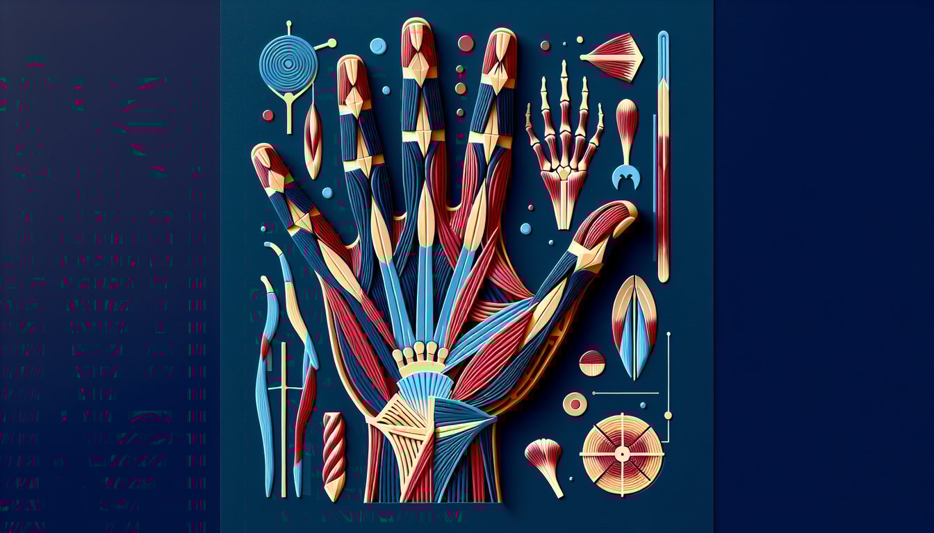
Use this Muscles of the Hand Quiz to check muscle names, actions, and functions, and see where you need review. For more practice, try a hand muscles quiz, build context with a forearm muscles quiz, or test bone landmarks in a carpal bones quiz as well.
Study Outcomes
- Identify Hand and Wrist Muscles -
After the muscles of the hand quiz, you'll be able to pinpoint major intrinsic and extrinsic hand and wrist muscles by name and anatomical position.
- Differentiate Muscle Functions -
Distinguish between flexors, extensors, and intrinsic muscles, understanding how each contributes to gripping, extension, and fine motor control.
- Locate Origins and Insertions -
Using this hand anatomy quiz, map out where key muscles originate and insert, improving your spatial awareness of the anatomy of the hand.
- Apply Anatomy to Movement -
Translate your knowledge of hand and wrist muscles into practical insights about grip mechanics and functional movement patterns.
- Evaluate Anatomical Knowledge -
Assess your current understanding of hand anatomy and pinpoint areas for further study to solidify your expertise.
- Retain Muscle Names and Actions -
Reinforce your memory of muscle names, locations, and primary actions, boosting confidence for academic or professional discussions.
Cheat Sheet
- Intrinsic vs. Extrinsic Hand Muscles -
Intrinsic muscles originate within the hand itself and govern fine motor movements like precision pinch, while extrinsic muscles start in the forearm and drive powerful grip and gross finger motion. Detailed in Gray's Anatomy and university courses, this classification is essential for your muscles of the hand quiz and clarifies the relationships between hand and wrist muscles.
- Flexor Digitorum Superficialis vs. Profundus -
The FDS tendon splits to allow the FDP tendon to pass through, flexing the PIP and DIP joints respectively. A quick mnemonic is "Superficialis = Superficial layer = PIP flexion," "Profundus = Deep layer = DIP flexion," as highlighted in the Journal of Hand Surgery.
- Extensor Compartments and Mnemonic -
The dorsal wrist contains six extensor compartments; memorize "Some Lovers Try Positions That They Can't Handle" (APL/EPB, EPL, ECRL/ECRB, EDC/EI, EDM, ECU) to recall tendon pathways. This handy trick from OpenStax Anatomy & Physiology clarifies which muscle extends which digit.
- Power vs. Precision Grips -
Power grips use extrinsic flexors like FDS/FDP and the intrinsic adductor pollicis for strong, forceful holds, while precision grips rely on lumbricals and interossei for delicate tasks. Studies in the Journal of Biomechanics show that key pinch (lateral pinch) predominantly uses adductor pollicis and the first dorsal interosseous muscle.
- Innervation and Clinical Correlation -
The median nerve supplies the thenar muscles and two lateral lumbricals (LOAF mnemonic), the ulnar nerve innervates the interossei, adductor pollicis, and medial lumbricals, and the radial nerve controls wrist and finger extensors. Remember "LOAF for median" to excel in the anatomy of the hand quiz and to predict deficits in carpal tunnel syndrome or ulnar neuropathies (American Society for Surgery of the Hand).