Unlock hundreds more features
Save your Quiz to the Dashboard
View and Export Results
Use AI to Create Quizzes and Analyse Results

Sign inSign in with Facebook
Sign inSign in with Google

Upper Limb Anatomy Quiz: Flex Your Knowledge!

Test Your Skills in Upper Limb Labeling & Muscle Anatomy!

Difficulty: Moderate
2-5mins
Learning OutcomesCheat Sheet
Paper cut style upper limb illustration showing arm bones muscles and joints on golden yellow background for anatomy quiz

This upper limb anatomy quiz helps you practice bones, muscles, and joints across the shoulder, arm, forearm, and hand. Work through quick questions to see what to review before an exam; for extra practice, try our upper extremity practice or start with the bones quiz .

Which bone forms the lateral border of the forearm?
Humerus
Radius
Scapula
Ulna
The radius is the lateral bone in the forearm when in the anatomical position, situated on the thumb side. The ulna lies medially alongside the little finger. The humerus is the upper arm bone, and the scapula is part of the shoulder girdle. For further reading, see .
What is the primary function of the deltoid muscle?
Protraction of the scapula
Flexion of the elbow
Abduction of the arm at the shoulder
Extension of the wrist
The deltoid muscle is the main abductor of the arm at the glenohumeral joint, particularly effective beyond the first 15 degrees. It also assists in flexion, extension, and rotation of the shoulder depending on which fibers are activated. It does not act on the elbow or wrist. More details at .
Which joint is formed by the articulation of the head of the humerus and the glenoid cavity of the scapula?
Humeroulnar joint
Sternoclavicular joint
Glenohumeral joint
Acromioclavicular joint
The glenohumeral joint, commonly known as the shoulder joint, is formed by the head of the humerus fitting into the glenoid cavity of the scapula, allowing a wide range of motion. The acromioclavicular joint connects the clavicle to the scapula's acromion process. The sternoclavicular joint links the clavicle to the sternum. For more, see .
The anatomical snuffbox is located on which side of the wrist?
Dorsal radial surface
Dorsal ulnar surface
Palmar radial surface
Palmar ulnar surface
The anatomical snuffbox is a triangular depression on the dorsal aspect of the wrist at the base of the thumb, bordered by the tendons of extensor pollicis longus and brevis. It is important clinically for palpating the scaphoid bone. It is not found on the palmar or ulnar sides. See for details.
Which muscle is the primary flexor of the wrist on the radial side?
Extensor carpi ulnaris
Pronator teres
Supinator
Flexor carpi radialis
Flexor carpi radialis flexes and abducts the wrist on the radial side. Extensor carpi ulnaris extends and adducts the wrist. Pronator teres pronates the forearm, and supinator supinates it. For more, see .
Which nerve innervates the triceps brachii muscle?
Musculocutaneous nerve
Ulnar nerve
Radial nerve
Median nerve
The radial nerve innervates all muscles in the posterior compartment of the arm, including the triceps brachii, responsible for elbow extension. The median and ulnar nerves serve anterior compartment muscles of the forearm. The musculocutaneous nerve supplies anterior arm flexors. See for more.
Which structure passes through the carpal tunnel?
Median nerve
Ulnar nerve
Flexor carpi ulnaris tendon
Radial artery
The carpal tunnel contains the flexor digitorum tendons, flexor pollicis longus tendon, and the median nerve. The ulnar nerve and artery pass outside the tunnel through Guyon's canal. The radial artery is also superficial, not within the tunnel. More at .
The clavicle articulates medially with which part of the axial skeleton?
Acromion process of the scapula
Coracoid process of the scapula
Scapular spine
Manubrium of the sternum
The medial (sternal) end of the clavicle articulates with the manubrium to form the sternoclavicular joint, the only bony articulation between the upper limb and axial skeleton. The acromion and coracoid processes are lateral scapular landmarks. For further reading, visit .
Beyond the lower border of teres major, the axillary artery continues as the __________.
Ulnar artery
Subclavian artery
Radial artery
Brachial artery
The axillary artery becomes the brachial artery once it passes the inferior border of the teres major muscle. The subclavian artery is proximal, and the radial and ulnar arteries are distal branches of the brachial. More details at .
Which rotator cuff muscle is most commonly torn in rotator cuff injuries?
Supraspinatus
Teres minor
Infraspinatus
Subscapularis
The supraspinatus tendon is the most frequently injured part of the rotator cuff due to its anatomical position under the acromion and its critical role in initiating arm abduction. Infraspinatus, teres minor, and subscapularis tears are less common. See .
At the elbow, the ulnar nerve lies superficial in a groove between which two structures?
Lateral epicondyle and radial head
Lateral epicondyle and olecranon
Medial epicondyle and olecranon
Medial epicondyle and trochlea
The ulnar nerve runs in the cubital tunnel, a groove between the medial epicondyle of the humerus and the olecranon of the ulna, making it vulnerable to compression and impact ('funny bone'). It does not lie near the lateral epicondyle. For more information, see .
Which ligament encircles the head of the radius and holds it against the ulna?
Ulnar collateral ligament
Radial collateral ligament
Interosseous ligament
Annular ligament
The annular ligament wraps around the circumference of the radial head, securing it to the radial notch of the ulna and allowing pronation and supination. Ulnar and radial collateral ligaments stabilize the elbow laterally, and the interosseous membrane connects shafts of radius and ulna. More at .
Which forearm muscle is innervated by both the median and ulnar nerves?
Pronator teres
Flexor carpi ulnaris
Flexor digitorum profundus
Supinator
The flexor digitorum profundus has dual innervation: the lateral half (digits 2 - 3) by the anterior interosseous branch of the median nerve and the medial half (digits 4 - 5) by the ulnar nerve. Pronator teres and supinator are solely innervated by the median and radial nerves respectively. See .
A fracture at the surgical neck of the humerus most likely damages which nerve and accompanying artery?
Median nerve and brachial artery
Axillary nerve and posterior circumflex humeral artery
Musculocutaneous nerve and brachial artery
Radial nerve and profunda brachii artery
The axillary nerve and the posterior circumflex humeral artery wrap around the surgical neck of the humerus, making them vulnerable in fractures at this site. The radial nerve and profunda brachii travel in the radial groove more distally, while the musculocutaneous and median nerves lie in the anterior compartment. More details at .
0
{"name":"Which bone forms the lateral border of the forearm?", "url":"https://www.quiz-maker.com/QPREVIEW","txt":"Which bone forms the lateral border of the forearm?, What is the primary function of the deltoid muscle?, Which joint is formed by the articulation of the head of the humerus and the glenoid cavity of the scapula?","img":"https://www.quiz-maker.com/3012/images/ogquiz.png"}

Study Outcomes

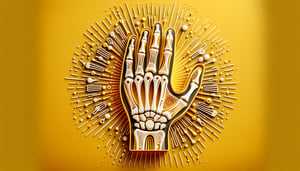
  1. Identify Major Bones of the Upper Limb -

    Accurately locate and name the principal bones of the upper extremity, including the humerus, radius, ulna, scapula, and clavicle, solidifying your understanding for the Ultimate Upper Limb Anatomy Quiz.
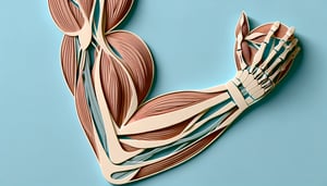
  2. Label Key Muscles -

    Recognize and label the primary muscles of the shoulder, arm, forearm, and hand, enhancing your performance in the upper limb muscles quiz.

  3. Differentiate Joint Structures -

    Distinguish between major articulations - such as the glenohumeral, elbow, and wrist joints - improving your comprehension of upper extremity anatomy quiz topics.

  4. Analyze Humerus Anatomy -

    Examine the anatomical landmarks and functional roles of the humerus, preparing you for detailed questions in the anatomy of the humerus quiz.

  5. Apply Anatomical Knowledge -

    Use your understanding of bones, muscles, and joints to solve clinical and practical scenarios, demonstrating mastery through the upper limb anatomy quiz challenge.

Cheat Sheet

  1. Shoulder Girdle Articulations -

    The shoulder girdle comprises the sternoclavicular, acromioclavicular, and glenohumeral joints - remember them in SAG order (Sterno-Acromio-Gleno). Each joint allows unique motion; the sternoclavicular joint enables clavicular rotations, and the glenohumeral joint provides the greatest range of motion in the upper extremity. Reviewing these in your upper limb anatomy quiz helps you identify bony landmarks and joint mechanics.

  2. Rotator Cuff Musculature (SITS) -

    The SITS muscles - Supraspinatus, Infraspinatus, Teres minor, Subscapularis - stabilize the glenohumeral joint and control rotation and abduction. A handy mnemonic is "SITS" to recall their sequence from superior to inferior; supraspinatus initiates abduction and infraspinatus/teres minor handle external rotation. Testing your knowledge of these muscles is crucial for the upper limb muscles quiz segment.

  3. Humerus Landmarks and Nerve Pathways -

    Key landmarks like the radial groove (home to the radial nerve) and the medial epicondyle (ulnar nerve's "funny bone") are vital for understanding nerve injury patterns. For example, a midshaft humeral fracture often endangers the radial nerve, leading to wrist drop. Integrate this into your upper limb anatomy quiz to link structure to clinical relevance.

  4. Forearm Muscle Compartments -

    The forearm divides into anterior (flexor-pronator) and posterior (extensor-supinator) compartments, primarily innervated by the median and radial nerves, respectively. Use the phrase "Flexors in Front" to recall that wrist and finger flexion muscles lie anteriorly, while extensors are - figuratively - "hitting the deck" posteriorly. This tip is perfect for mastering the upper limb labeling challenges in your quiz.

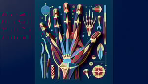
  5. Hand Intrinsics and Arch Mechanics -

    The hand's functional arches - longitudinal, proximal transverse, distal transverse - facilitate grip precision, supported by lumbricals and interossei. Remember "PAD & DAB": Palmar interossei ADduct, Dorsal interossei ABduct. Including this in your upper limb anatomy quiz ensures you can label structures and describe their roles in hand movements.

Powered by: Quiz Maker