Nail Anatomy Quiz: How Nails Grow and What Each Part Does
Quick, free nail structure quiz. Instant results with simple explanations.
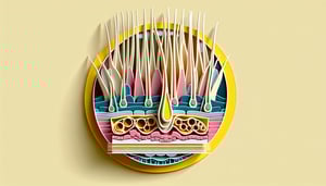
This nail anatomy quiz helps you check how nails grow and what each part does, from the matrix to the nail bed. Get quick questions with instant feedback, then build on your knowledge with our nail quiz for beginners, the hand bones quiz, or a hair anatomy quiz.
Study Outcomes
- Identify the Nail Growth Region -
Pinpoint the matrix as this region of the nail responsible for nail growth and describe how it produces new nail cells.
- Understand Key Nail Anatomy Components -
Distinguish between the nail bed, cuticle, lunula, and other parts to map out overall nail structure.
- Recall Nail Growth Facts -
Memorize essential nail growth facts such as average growth rates and factors influencing healthy nail development.
- Analyze Nail Structure Trivia -
Evaluate trivia questions to deepen your insight into how different nail regions interact and support growth.
- Apply Nail Health Self-Assessment -
Use targeted quiz questions to assess your nail health and identify when to seek professional care.
Cheat Sheet
- Nail Matrix: Growth Powerhouse -
The nail matrix, located under the proximal nail fold, is this region of the nail responsible for nail growth; its proliferating keratinocytes generate new nail plate cells through mitosis. Think "M for Matrix = Make more cells" to remember its role (Gray's Anatomy, 2021).
- Lunula: Matrix Visibility Clue -
The lunula is the visible whitish half-moon at the nail's base, reflecting active matrix function; a large lunula often indicates robust growth, while shape or color changes may signal health concerns. This concept often pops up in a nail anatomy quiz to test recognition of growth zones.
- Keratinization in the Nail Plate -
As matrix cells harden, they undergo keratinization to form the nail plate - a durable structure of fibrous proteins. Mnemonic "K.N.P. = Keratinization Creates Nail Plate" helps lock in key nail growth facts from dermatology references (NCBI).
- Growth Rate & Influencing Factors -
Average fingernails grow about 3.5 mm per month and toenails about 1.5 mm per month; age, nutrition, hormonal shifts, and systemic disease can accelerate or slow this rate (American Academy of Dermatology). Use Growth (mm) = Rate × Time to estimate expected nail length over weeks.
- Protective Nail Folds & Cuticle -
The proximal and lateral nail folds plus the eponychium (cuticle) shield the matrix from pathogens and moisture loss, essential for steady growth; think "folds hold and protect the gold (matrix)." In nail structure trivia and nail health questions, a damaged cuticle often precedes growth irregularities.