Unlock hundreds more features
Save your Quiz to the Dashboard
View and Export Results
Use AI to Create Quizzes and Analyse Results

Sign inSign in with Facebook
Sign inSign in with Google

Nervous System Multiple Choice Quiz - Test Your Knowledge

Think you can ace this mcq on the nervous system? Start now!

Difficulty: Moderate
2-5mins
Learning OutcomesCheat Sheet
Paper art brain with neuron branches and quiz prompts on dark blue background for a multiple choice nervous system challenge

This nervous system quiz helps you practice key ideas with 10 multiple-choice questions and instant feedback. Use it to spot gaps before your exam and strengthen recall of neurons, reflexes, and brain regions. For more practice, explore more practice questions .

What is the primary functional unit of the nervous system?
Glial cell
Neuron
Synapse
Axon
Neurons are the basic structural and functional units of the nervous system. They are specialized for transmitting electrical and chemical signals throughout the body. Each neuron consists of dendrites to receive input, a cell body to process signals, and an axon to transmit output. This enables rapid and precise communication between different parts of the body. For more information, see .
Which part of a neuron typically receives incoming signals from other neurons?
Dendrites
Cell body (soma)
Synaptic cleft
Axon
Dendrites are branched extensions of the neuron that receive electrical and chemical signals from other neurons. They convey these signals to the cell body for processing. Without dendrites, neurons would not be able to integrate information from multiple sources. For more details, see .
What structure connects the left and right cerebral hemispheres and facilitates interhemispheric communication?
Corpus callosum
Longitudinal fissure
Central sulcus
Fornix
The corpus callosum is a large band of white matter composed of axonal fibers that connects the two cerebral hemispheres. It allows communication and coordination between the left and right sides of the brain. Damage to this structure can impair interhemispheric transfer of information. Learn more at .
Which neurotransmitter is the primary inhibitory neurotransmitter in the adult mammalian central nervous system?
Glutamate
Dopamine
GABA (gamma-aminobutyric acid)
Acetylcholine
GABA is the chief inhibitory neurotransmitter in the adult mammalian brain, reducing neuronal excitability throughout the nervous system. It binds to GABA receptors, causing hyperpolarization of the postsynaptic membrane. Without sufficient GABA activity, neural circuits may become overactive and contribute to seizures. More information at .
The blood-brain barrier is mainly formed by which of the following cellular structures?
Ependymal cilia
Astrocytic endfeet
Tight junctions between endothelial cells
Microglia
The blood-brain barrier is created by tight junctions between endothelial cells lining the brain's capillaries, preventing many substances from crossing into the brain tissue. Astrocytes support and regulate the barrier but do not form the junctions themselves. This barrier protects neural tissue from toxins and pathogens. For more details, visit .
Which glial cell type is responsible for myelinating axons in the peripheral nervous system?
Oligodendrocytes
Schwann cells
Astrocytes
Microglia
Schwann cells are the glial cells in the peripheral nervous system that wrap around axons to form the myelin sheath, increasing the speed of electrical conduction. In contrast, oligodendrocytes perform this function in the central nervous system. Damage to Schwann cells can lead to peripheral neuropathies. Learn more at .
Which region of the brain is primarily responsible for coordination and fine-tuning of voluntary movement and balance?
Cerebellum
Medulla oblongata
Thalamus
Basal ganglia
The cerebellum integrates sensory input and motor commands to fine-tune and coordinate voluntary movements and maintain balance and posture. It receives input from proprioceptive sensors and the cerebral cortex to modulate motor output. Lesions in the cerebellum often lead to ataxia and coordination deficits. More at .
What describes an excitatory postsynaptic potential (EPSP)?
A transient depolarization that increases the likelihood of action potential generation
A transient hyperpolarization that decreases the likelihood of action potential generation
A sustained depolarization leading directly to multiple action potentials
A release of neurotransmitter into the synaptic cleft
An EPSP is a brief depolarization of the postsynaptic membrane potential, making the neuron more likely to fire an action potential. It results from the opening of ligand-gated ion channels that allow positive ions to enter. Unlike a hyperpolarizing IPSP, an EPSP moves the membrane potential closer to threshold. For more details, see .
Afferent neurons carry sensory information in which direction?
Within interneuron circuits in the spinal cord exclusively
Between different brain regions only
From peripheral receptors toward the central nervous system
From the central nervous system to muscles and glands
Afferent (sensory) neurons transmit sensory signals from peripheral receptors toward the central nervous system. Efferent neurons, in contrast, carry motor commands away from the CNS. Interneurons connect neurons within the CNS but are neither strictly afferent nor efferent. Learn more at .
Broca's area, critical for speech production, is located in which lobe of the cerebral cortex?
Right parietal lobe
Occipital lobe
Left temporal lobe
Left frontal lobe
Broca's area is located in the posteroinferior region of the frontal lobe, usually in the left hemisphere, and is essential for speech production and language processing. Damage to this area can result in expressive aphasia, characterized by difficulty in forming words. It works in concert with Wernicke's area via neural pathways for fluent communication. More information at .
0
{"name":"What is the primary functional unit of the nervous system?", "url":"https://www.quiz-maker.com/QPREVIEW","txt":"What is the primary functional unit of the nervous system?, Which part of a neuron typically receives incoming signals from other neurons?, What structure connects the left and right cerebral hemispheres and facilitates interhemispheric communication?","img":"https://www.quiz-maker.com/3012/images/ogquiz.png"}

Study Outcomes

  1. Identify Nervous System Components -

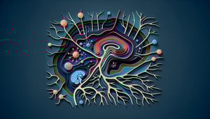
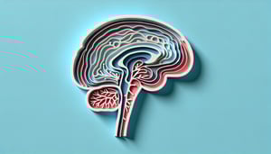
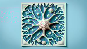
    Recognize major structures such as neurons, synapses, and neural pathways to solidify foundational knowledge of nervous system anatomy and physiology.

  2. Differentiate Neuron Types and Functions -

    Explain the roles of sensory, motor, and interneurons in signal transmission and interpret how synaptic interactions influence neural communication.

  3. Apply Knowledge Through MCQs -

    Use your understanding to tackle nervous system multiple choice questions with answers, reinforcing concepts with immediate feedback.

  4. Analyze Reflex and Signal Transmission -

    Break down the steps involved in reflex arcs and nerve impulse propagation to enhance comprehension of neurophysiological processes.

  5. Interpret Quiz Feedback -

    Review explanations for correct and incorrect answers to identify knowledge gaps and strengthen retention of key facts.

  6. Evaluate Learning Progress -

    Track your quiz score to monitor improvement, set study goals, and focus on areas needing further review.

Cheat Sheet

  1. Neuron Anatomy and Resting Potential -

    Neurons consist of a soma, dendrites, and an axon that work together to transmit electrical signals. The resting membrane potential (~ - 70 mV) arises from K+ leak channels and the Na+/K+ ATPase pump, as described by the Goldman equation. Mnemonic: "PEK" helps recall that Potassium Equilibrium sets the resting membrane Potential (NIH).

  2. Action Potential Dynamics -

    Action potentials follow an "all-or-none" law and proceed through depolarization, repolarization, and hyperpolarization phases. Voltage-gated Na+ channels open rapidly, then inactivate, while K+ channels restore the membrane potential (Nature Reviews Neuroscience). Remember "D-R-H" for Depolarize, Repolarize, Hyperpolarize.

  3. Synaptic Transmission and Neurotransmitters -

    Neurotransmitter release at the synaptic cleft is Ca2+-dependent and can be excitatory (e.g., glutamate) or inhibitory (e.g., GABA) as detailed by Oxford University research. Synaptic strength is modulated by receptor density and reuptake mechanisms, which MCQs often test. Example: Acetylcholine (ACh) at the neuromuscular junction triggers muscle contraction.

  4. Autonomic Nervous System Balances -

    The autonomic system divides into sympathetic (fight-or-flight) and parasympathetic (rest-and-digest) branches, each using distinct neurotransmitters (norepinephrine vs. ACh). Understanding antagonistic effects - like pupil dilation versus constriction - is a common MCQ topic (American Physiological Society). Use "SLUDD" for Parasympathetic: Salivation, Lacrimation, Urination, Digestion, Defecation.

  5. Reflex Arcs and Neural Circuits -

    Simple reflex arcs involve a sensory receptor, afferent neuron, integration center, efferent neuron, and effector - no brain involvement necessary (NCBI). Polysynaptic reflexes add interneurons, while higher centers (cortex, cerebellum) modify responses. Practice drawing the knee-jerk reflex pathway to solidify this concept.

Powered by: Quiz Maker