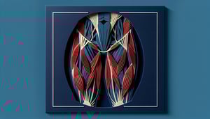
Pelvic Girdle Quiz: Bones and Lower Limb Anatomy
Quick, free lower limb anatomy quiz to check your recall. Instant results.
This quiz helps you review the pelvic girdle and lower limb anatomy, covering bones, landmarks, and joints. For extra practice, see the pelvis labeling quiz, try the os coxae labeling quiz, or work on muscles in the leg muscles quiz. Instant feedback highlights what to revisit before class or lab.
Study Outcomes
- Identify Major Bones -
Identify and label the pelvic girdle and lower limb bones, including the ilium, ischium, femur, tibia, and fibula, with accuracy.
- Describe Hip Joint Anatomy -
Describe the structural components of the hip joint, including the acetabulum, femoral head, and supporting ligaments, and explain their functional roles.
- Distinguish Pelvic Girdle Muscles -
Distinguish the primary muscles of the pelvic girdle such as the gluteal group and iliopsoas by origin, insertion, and action.
- Analyze Lower Limb Bone Landmarks -
Analyze key landmarks on the femur, tibia, fibula, and foot bones to understand their clinical and biomechanical significance.
- Apply Anatomical Knowledge -
Apply understanding of lower limb bones and hip joint anatomy to clinical scenarios, such as common fracture sites and injury mechanisms.
- Recall Functional Movements -
Recall and describe the major movements - flexion, extension, abduction, adduction, and rotation - of the hip and lower limb joints as tested in the pelvic girdle and lower limb anatomy quiz.
Cheat Sheet
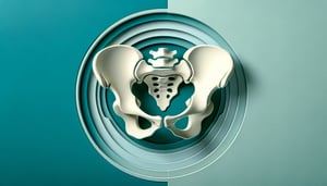
- Pelvic Girdle Overview -
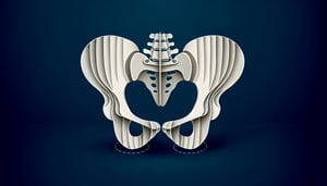
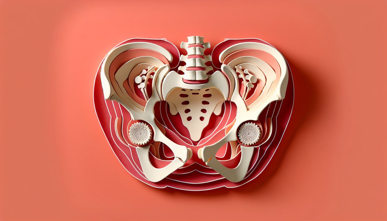
The pelvic girdle comprises the ilium, ischium, and pubis, fused at the acetabulum to articulate with the femur. It supports abdominal organs and transfers weight from the axial skeleton to the lower limbs. Remember "IPA" (Ilium, Pubis, Ischium) to recall its three bones quickly.
- Hip Joint Anatomy -
The hip joint is a ball-and-socket synovial joint formed by the femoral head and the acetabulum, offering flexion, extension, abduction, adduction, and rotation. Strong ligaments (iliofemoral, pubofemoral, and ischiofemoral) reinforce stability - iliofemoral is the Y-shaped "bigelow" ligament. When quizzing yourself, visualize the femoral head "nestled" in the acetabular cup like a golf ball in a tee.
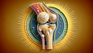
- Key Femur and Tibia Landmarks -
The femur's head, neck, greater trochanter, and linea aspera are critical for muscle attachments; the tibia's plateau and tuberosity guide knee mechanics. Clinically, the femoral neck's angle of inclination (~125°) affects gait and is measured in anterior - posterior X-rays. Use the mnemonic "He Never Tried Lacing Shoes" (Head, Neck, Trochanter, Linea Aspera, Shaft) to recall femoral landmarks.
- Pelvic Girdle Muscles -
Major muscles - gluteus maximus, medius, minimus, and the lateral rotators - stabilize the pelvis during stance and control hip motion. The gluteus medius is the "workhorse" for hip abduction and prevents contralateral hip drop (Trendelenburg sign). For memorization, think "Max, Med, Min" stacking from superficial to deep.
- Foot Arches and Lower Limb Bones -
The medial longitudinal, lateral longitudinal, and transverse arches of the foot absorb shock and distribute body weight during locomotion. Key bones include the tarsals (e.g., talus and calcaneus), metatarsals, and phalanges; weak arches can lead to flatfoot. Remember "Tom, Dick And Very Nervous Harry" for tarsal names and practice identifying them in radiographic images.