Unlock hundreds more features
Save your Quiz to the Dashboard
View and Export Results
Use AI to Create Quizzes and Analyse Results

Sign inSign in with Facebook
Sign inSign in with Google

How Well Do You Know the Cup-Like Collecting Region of the Renal Pelvis?

Ready to ace renal pelvis anatomy? Try this collecting region quiz now!

Difficulty: Moderate
2-5mins
Learning OutcomesCheat Sheet
Paper art illustration for renal pelvis anatomy quiz on a teal background

This Renal Pelvis Quiz helps you review the renal pelvis and the kidney's cup-like collecting region, from minor to major calyces, so you can practice key anatomy fast. Use it to spot gaps before an exam, then try the pelvis practice or the urinary system practice for more review.

The funnel-shaped upper end of the ureter within the kidney that serves as the initial collecting area for urine is known as what?
Renal pelvis
Minor calyx
Renal papilla
Renal cortex
The renal pelvis is the funnel-shaped expansion at the upper end of the ureter inside the kidney, acting as a reservoir for urine before it enters the ureter. It collects urine from the major calyces, while the renal cortex is the outer parenchyma involved in filtration. Minor calyces and papillae are smaller collecting structures upstream of the pelvis.
The renal pelvis receives urine directly from which of the following structures?
Proximal convoluted tubules
Major calyces
Minor calyces
Renal artery
Urine flows from the collecting ducts into minor calyces, which converge into major calyces; these then empty directly into the renal pelvis. The proximal convoluted tubule and renal artery are unrelated to this collecting sequence.
Which type of epithelium lines the inner surface of the renal pelvis?
Pseudostratified columnar epithelium
Stratified squamous epithelium
Simple cuboidal epithelium
Transitional epithelium
The renal pelvis is lined by transitional epithelium (urothelium), which can stretch to accommodate varying volumes of urine. Simple cuboidal, stratified squamous, and pseudostratified columnar epithelia are found in other regions of the urinary or respiratory tracts.
Within the kidney, the renal pelvis is located in which region?
Renal cortex
Fibrous capsule
Renal sinus
Renal medulla
The renal pelvis resides in the renal sinus, a fat-filled cavity at the hilum of the kidney that also contains vessels and nerves. The cortex and medulla are parenchymal regions involved in filtration and urine concentration. The fibrous capsule is the outer covering.
Typically, how many major calyces converge to form a single renal pelvis in a human kidney?
Two
Four
One
Three
Most human kidneys have two major calyces (superior and inferior) that join to form the renal pelvis. While variations exist, a single calyx or more than two are uncommon.
Which mechanism propels urine from the renal pelvis into the ureter?
Osmotic flow
Peristalsis of smooth muscle
Glomerular filtration
Passive diffusion
Smooth muscle in the walls of the renal pelvis and ureter generates peristaltic waves to move urine downward. Filtration occurs in the glomerulus, and passive diffusion or osmosis do not drive bulk urine flow.
Which imaging technique is classically used to visualize the anatomy of the renal pelvis and ureters?
MRI of the brain
Intravenous pyelogram
Ultrasound of the neck
CT scan of the chest
An intravenous pyelogram (IVP) involves injecting contrast dye and taking serial X-rays, highlighting the collecting system including the renal pelvis. Other modalities target unrelated regions.
Obstruction of urine flow in the renal pelvis commonly leads to which condition?
Hydronephrosis
Renal agenesis
Glycosuria
Nephritis
Hydronephrosis describes dilation of the renal pelvis and calyces due to obstruction of urinary outflow. Nephritis is inflammation, glycosuria refers to glucose in urine, and renal agenesis is absence of a kidney.
The renal pelvis directly drains into which structure?
Ureter
Minor calyx
Collecting duct
Glomerulus
Urine travels from the renal pelvis into the ureter, which conveys it to the urinary bladder. Collecting ducts empty into minor calyces, and glomeruli are filtration units upstream.
The indentation of the kidney where the renal vessels and ureter enter and exit is called the what?
Renal papilla
Renal capsule
Renal hilum
Medullary ray
The renal hilum is the concave region on the kidney's medial side where the renal artery, vein, and ureter pass. Papillae are the tips of medullary pyramids. Medullary rays are radial striations in the cortex.
The luminal surface of the renal pelvis exhibits which feature to accommodate changes in urine volume?
Rugae folds
Villi
Cilia
Microvilli
Rugae are mucosal folds that allow the transitional epithelium of the renal pelvis to stretch as urine volume fluctuates. Villi and microvilli increase surface area for absorption in the gut and renal tubules, respectively, and cilia are absent here.
Which artery provides the primary blood supply to the renal pelvis?
Inferior phrenic artery
Superior mesenteric artery
Branches of the renal artery
Internal iliac artery
The renal pelvis receives arterial blood from branches of the renal artery, specifically segmental, interlobar, and arcuate arteries. The other listed arteries supply the diaphragm, gut, and pelvic organs.
A typical human kidney contains how many minor calyces?
20 to 30
1 to 2
3 to 5
8 to 18
Minor calyces are cup-shaped structures that receive urine from renal papillae; a normal human kidney has about 8 to 18 of them. Fewer than 8 or more than 18 are rare anatomical variations.
The anatomical junction between the renal pelvis and the proximal ureter is called the what?
Ureterovesical junction
Pelviureteric junction
Vesicoureteric junction
Renal papillary junction
The pelviureteric junction (PUJ) marks the transition from the renal pelvis into the ureter. The vesicoureteric or ureterovesical junction lies between the ureter and bladder.
Lymphatic drainage from the renal pelvis primarily goes to which lymph node group?
Iliac nodes
Lateral aortic (lumbar) nodes
Inguinal nodes
Para-rectal nodes
Lymphatics from the renal pelvis and surrounding kidney parenchyma drain into the lateral aortic (lumbar) lymph nodes located near the abdominal aorta. The other nodal groups are associated with lower limb or pelvic drainage.
Which nerve plexus provides autonomic innervation to the renal pelvis?
Renal plexus
Hypogastric plexus
Pelvic plexus
Celiac plexus
The renal plexus, deriving sympathetic fibers from the lesser splanchnic nerves (T10 - T12) and parasympathetic fibers from the vagus nerve, innervates the renal pelvis. The pelvic and hypogastric plexuses supply pelvic organs.
On ultrasound, an anteroposterior pelvic diameter exceeding which measurement in adults suggests dilation?
8 mm
10 mm
20 mm
4 mm
An anteroposterior diameter of the renal pelvis over 10 mm on ultrasound is considered hydronephrosis in adults. Measurements under this threshold are generally normal.
The renal pelvis is embryologically derived from which of the following structures?
Metanephric blastema
Ureteric bud
Mesonephric duct
Pronephric duct
The ureteric bud, an outgrowth of the mesonephric duct, gives rise to the ureters, renal pelvis, calyces, and collecting ducts. The metanephric blastema forms nephrons.
The muscular layer of the renal pelvis is composed of which type of muscle tissue?
Smooth muscle in circular and longitudinal layers
Striated skeletal muscle
Cardiac muscle
Smooth muscle only in a single layer
The wall of the renal pelvis has two layers of smooth muscle - inner longitudinal and outer circular - that contract rhythmically for peristalsis. Skeletal and cardiac muscle are not present.
Stones lodged at the pelviureteric junction most commonly cause referred pain in which dermatomal distribution?
L4 - S1
T11 - L2
C7 - T1
T4 - T6
Pain from ureteric obstruction at the pelviureteric junction is referred along the T11 - L2 dermatomes, often perceived in the flank or groin. Other dermatomes are associated with different regions.
In a retrograde pyelogram, contrast material is introduced through which route?
Arterial puncture
Bladder irrigation
Intravenous injection
Ureter via cystoscopic catheter
A retrograde pyelogram involves passing a catheter through the bladder and ureteral orifice to inject contrast directly into the ureter and pelvicalyceal system. Intravenous techniques describe an IVP.
In hydronephrosis, which structure dilates first as urine backs up?
Loop of Henle
Proximal tubule
Renal pelvis
Glomerulus
When urinary outflow is obstructed, the renal pelvis dilates initially, followed by calyces and eventually the parenchyma. The filtration and tubular structures are affected later.
Which congenital anomaly at the pelviureteric junction can cause chronic obstructive uropathy?
PUJ obstruction
Ectopic kidney
Horseshoe kidney
Double ureter
Pelviureteric junction (PUJ) obstruction is a congenital narrowing where the pelvis meets the ureter, leading to hydronephrosis. Double ureter and other anomalies may affect drainage but are distinct entities.
The basement membrane of the transitional epithelium in the renal pelvis is primarily composed of which collagen type?
Type I collagen
Type II collagen
Type IV collagen
Type III collagen
Basement membranes, including those under urothelium in the renal pelvis, are rich in type IV collagen, providing a scaffold for cell attachment. Types I and III are found in interstitial connective tissues, while type II is in cartilage.
Sympathetic fibers supplying the renal pelvis originate from which spinal segments via the lesser splanchnic nerve?
T10 - T11
S2 - S4
T1 - T4
L2 - L4
The lesser splanchnic nerve arising from T10 - T11 spinal segments contributes sympathetic fibers to the renal plexus, innervating the renal pelvis. Other segmental levels supply different organs.
Urothelial cells lining the renal pelvis commonly express which immunohistochemical marker?
CD10
S100
WT1
Uroplakin III
Uroplakin III is a transmembrane protein specific to urothelial cells, including those in the renal pelvis, and is used as a diagnostic marker. CD10 and WT1 are found in renal tubule and podocyte lineages, respectively.
Which embryologic event gives rise to the renal pelvis during kidney development?
Invagination of the glomerular capsule
Branching of the ureteric bud
Fusion of mesonephric ducts
Differentiation of metanephric blastema
Branching of the ureteric bud forms the collecting system, including the renal pelvis, calyces, and collecting ducts. The metanephric blastema differentiates into nephrons, while glomerular capsule invagination forms glomeruli.
A ureter that passes posterior to the inferior vena cava, causing pelviureteric obstruction, is termed what anomaly?
Retrocaval ureter
Duplex collecting system
Pelvic kidney
Congenital PUJ obstruction
A retrocaval (or circumcaval) ureter deviates behind the IVC, leading to compression and hydronephrosis. Duplex systems involve duplicated ureters, and pelvic kidneys are ectopic.
Which genetic mutation is most commonly associated with upper tract urothelial carcinoma affecting the renal pelvis?
NF1 mutation
BRCA1 mutation
FGFR3 mutation
JAK2 mutation
Activating mutations in FGFR3 are frequent in urothelial carcinomas of the renal pelvis and bladder. BRCA1, NF1, and JAK2 are linked to other neoplasms.
In diuretic renography, retention of tracer in the renal pelvis beyond 20 minutes suggests what condition?
Normal renal function
Vesicoureteral reflux
Obstructive uropathy
Pyelonephritis
Delayed tracer clearance from the renal pelvis after diuretic administration indicates obstructive uropathy. Normal kidneys clear tracer rapidly, and pyelonephritis or reflux have different scintigraphic patterns.
Pacemaker cells similar to those in the gastrointestinal tract that coordinate peristalsis in the renal pelvis are known as?
Podocytes
Macula densa cells
Juxtaglomerular cells
Interstitial cells of Cajal-like cells
Cells resembling gastrointestinal interstitial cells of Cajal have been identified in the upper ureter and renal pelvis, acting as pacemakers for peristaltic contractions. The others are renal filtration or endocrine elements.
Hypercalciuria predisposes to which type of renal pelvis pathology?
Malignant transformation
Cystic dilation
Acute infection
Calcium stone formation
Excess urinary calcium (hypercalciuria) increases the risk of calcium-based kidney stones that often lodge in the renal pelvis. Cystic changes and malignancy have different etiologies.
A bifid renal pelvis describes which anatomic variant?
Complete double ureter
Pelvic kidney
Partial duplication of the pelvis
Horseshoe-shaped pelvis
A bifid pelvis has two branches that unite into a single ureter, representing partial duplication. Complete duplication yields two ureters. Horseshoe and pelvic kidneys are ectopic anomalies.
On CT urography, a normal renal pelvis axial diameter is typically less than how many millimeters?
20 mm
5 mm
15 mm
10 mm
A renal pelvis measuring under 5 mm in diameter on non-distended CT is considered normal. Measurements exceeding 10 - 15 mm often indicate dilation.
Which receptor subtype on the smooth muscle of the renal pelvis primarily mediates peristaltic contraction?
?2-adrenergic receptor
Histamine H1 receptor
?1-adrenergic receptor
Muscarinic M2 receptor
?1-adrenergic receptors on pelvic and ureteric smooth muscle modulate tone and peristaltic frequency. ?2, muscarinic, and histamine receptors play lesser roles in urinary tract peristalsis.
Genomic studies of urothelial carcinoma in the renal pelvis frequently reveal alterations in which signaling pathway?
Hedgehog pathway
PI3K/AKT pathway
TGF-? pathway
Notch pathway
Many urothelial carcinomas, including those in the renal pelvis, harbor mutations activating the PI3K/AKT signaling cascade, contributing to cell proliferation and survival. The other pathways are implicated in different cancer types.
Which combination of cytokeratins is characteristically expressed by urothelial cells of the renal pelvis?
CK20 and CK17
CK1 and CK10
CK8 and CK18
CK5 and CK15
Cytokeratins 8 and 18 are intermediate filament proteins expressed in simple epithelia, including transitional urothelium. CK20 is more superficial in bladder urothelium, and CK5/15 mark basal cells in stratified epithelia.
Basement membrane adhesion in the renal pelvis epithelium is mediated by which key extracellular matrix protein?
Collagen I
Laminin-5
Fibronectin
Elastin
Laminin-5, a heterotrimeric glycoprotein in the basement membrane, promotes adhesion of urothelial cells to underlying stroma. Elastin and collagen I provide elasticity and tensile strength in other tissues.
Studies of peristaltic dysfunction in pelviureteric junction obstruction implicate overexpression of which signaling molecule in smooth muscle hyperplasia?
EGF
VEGF
PDGF
TGF-?1
Transforming growth factor-?1 (TGF-?1) is upregulated in PUJ obstruction tissues, stimulating smooth muscle proliferation and fibrosis. EGF, VEGF, and PDGF have roles in other vascular and epithelial responses.
0
{"name":"The funnel-shaped upper end of the ureter within the kidney that serves as the initial collecting area for urine is known as what?", "url":"https://www.quiz-maker.com/QPREVIEW","txt":"The funnel-shaped upper end of the ureter within the kidney that serves as the initial collecting area for urine is known as what?, The renal pelvis receives urine directly from which of the following structures?, Which type of epithelium lines the inner surface of the renal pelvis?","img":"https://www.quiz-maker.com/3012/images/ogquiz.png"}

Study Outcomes

  1. Identify Renal Pelvis Components -

    Recognize and name the cup-like structures that form the collecting region of the renal pelvis, including major and minor calyces.

  2. Explain Urine Flow Pathways -

    Describe how urine travels from the kidney collecting ducts through the renal pelvis into the ureter.

  3. Differentiate Calyx Types -

    Distinguish between major and minor calyces based on their anatomical features and functions within renal pelvis anatomy.

  4. Analyze Structural Relationships -

    Examine how the cup-like collecting region interfaces with surrounding kidney tissue and the urinary system.

  5. Apply Anatomical Knowledge Clinically -

    Use your understanding of the renal pelvis to interpret common clinical scenarios and imaging of the urinary system.

  6. Recall Collecting Duct Organization -

    Memorize the arrangement of kidney collecting ducts as they converge into the renal pelvis for effective quiz performance.

Cheat Sheet

  1. Minor Calyces Structure -

    Each kidney typically has 8 - 18 minor calyces, each forming a neat cup around a single renal papilla to collect newly formed urine. Mnemonic: "MINor = Manifolds IN charge" helps recall that each minor calyx handles an INdividual papilla. (Source: Netter's Atlas of Human Anatomy)

  2. Major Calyces Convergence -

    Two or three minor calyces merge to form larger major calyces, which serve as intermediate cups before urine enters the renal pelvis. Remember the "2 - 3 Rule" for easy recall: 2 - 3 minor calyces per major calyx. (Source: Gray's Anatomy)
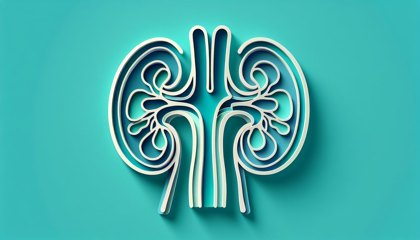
  3. Renal Pelvis Funnel -

    The renal pelvis is a funnel-shaped expansion of the proximal ureter lined by transitional epithelium, ensuring a watertight barrier to urine backflow. Think of it as the "grand central station" directing traffic from major calyces into the ureter. (Source: University of Michigan Medical School)

  4. Transitional Epithelium Function -

    Transitional (urothelial) cells in the calyces and pelvis stretch to accommodate varying urine volumes while preventing leakage or reabsorption. Use the formula "Elasticity + Impermeability = Urothelial Efficiency" to remember its dual role. (Source: Journal of Histology and Cytology)

  5. Clinical Significance in Nephrolithiasis -

    Kidney stones often lodge in the calyces' cup-like recesses, causing hydronephrosis upstream. On ultrasound or CT urogram, calyceal dilation signals obstruction - think "Cup Capture" for stone entrapment. (Source: American Urological Association)

Powered by: Quiz Maker