Unlock hundreds more features
Save your Quiz to the Dashboard
View and Export Results
Use AI to Create Quizzes and Analyse Results

Sign inSign in with Facebook
Sign inSign in with Google

Think You Know the Integumentary System? Take the Quiz!

Take our integumentary practice test and prove your skin savvy!

Difficulty: Moderate
2-5mins
Learning OutcomesCheat Sheet
Paper cut illustration of skin cross section with layers hair follicle sweat glands and quiz heading on teal background

This integumentary system quiz helps you practice skin anatomy and functions so you can spot gaps before your exam. Answer quick questions on layers, glands, hair, pigments, and healing, then use our skin layers practice for a fast, focused review.

Which layer is the outermost layer of the epidermis?
Stratum spinosum
Stratum granulosum
Stratum basale
Stratum corneum
The stratum corneum is the outermost layer of the epidermis, consisting of dead, flattened keratinocytes that form a protective barrier against pathogens and water loss. It is continuously shed and replaced by cells from deeper layers. This layer is essential for skin's barrier function.
Which cell type produces the pigment melanin?
Melanocyte
Keratinocyte
Fibroblast
Langerhans cell
Melanocytes are specialized cells in the stratum basale that synthesize melanin, the pigment responsible for skin and hair color. They transfer melanin granules to keratinocytes to protect nuclei from UV damage. Their activity determines individual pigmentation and response to sunlight.
The dermis is primarily composed of which type of tissue?
Adipose tissue
Epithelial tissue
Smooth muscle
Dense irregular connective tissue
The dermis is mainly made of dense irregular connective tissue containing collagen and elastin fibers, which provide strength and elasticity. It houses blood vessels, nerves, hair follicles, and glands. This arrangement allows the skin to withstand stretching in multiple directions.
The primary function of sebaceous glands is to secrete:
Sebum
Cerumen
Saliva
Sweat
Sebaceous glands secrete sebum, an oily substance that lubricates hair and skin, preventing dryness and providing a barrier against microbes. Sebum is produced via holocrine secretion when entire cells disintegrate to release lipids. Overactivity can lead to acne formation.
The hypodermis primarily stores:
Melanin
Fat
Keratin
Collagen
The hypodermis, or subcutaneous layer, is rich in adipose tissue that stores fat for energy, insulation, and cushioning. It anchors the skin to underlying structures and helps regulate body temperature. Its thickness varies by body region and individual.
Which receptor in the skin is most sensitive to light touch?
Ruffini ending
Pacinian corpuscle
Free nerve ending
Meissner's corpuscle
Meissner's corpuscles are rapidly adapting mechanoreceptors located in dermal papillae, best suited for detecting light touch and low-frequency vibrations. They are especially concentrated in fingertips and lips. Their structure allows for sensitivity to fine tactile stimuli.
Which layer of the epidermis is found only in thick skin?
Stratum corneum
Stratum granulosum
Stratum basale
Stratum lucidum
The stratum lucidum is a clear, thin layer of dead keratinocytes found only in thick skin areas such as the palms and soles. It lies between the stratum corneum and stratum granulosum and provides extra protection. This layer is absent in thin skin.
What is the main role of keratin in the skin?
Provide strength and water resistance
Produce pigment
Mediate immune response
Generate heat
Keratin is a fibrous protein produced by keratinocytes that provides mechanical strength and waterproofing to the skin. It forms intermediate filaments within cells, contributing to barrier function. Disorders of keratin can lead to skin fragility.
Eccrine sweat glands differ from apocrine sweat glands in that eccrine glands:
Begin functioning at puberty
Produce odorous secretions
Open into hair follicles
Secrete via merocrine secretion
Eccrine sweat glands release their watery secretions directly onto the skin surface via merocrine secretion without shedding cellular material. They play a key role in thermoregulation and are active from birth. Apocrine glands, in contrast, open into hair follicles and activate at puberty.
Which phase of wound healing is characterized by collagen deposition and scar formation?
Hemostasis
Proliferation phase
Remodeling phase
Inflammatory phase
The remodeling phase follows proliferation and involves collagen maturation and reorganization, leading to scar tissue formation. Type III collagen is replaced by stronger type I collagen, and the wound gains tensile strength. This phase can last for months.
Which cells in the epidermis act as antigen-presenting cells?
Melanocytes
Keratinocytes
Langerhans cells
Merkel cells
Langerhans cells are specialized dendritic cells in the stratum spinosum that capture and process antigens, then migrate to lymph nodes to initiate immune responses. They play a crucial role in skin immunosurveillance. Damage to these cells can impair cutaneous immunity.
Thick skin is found in all of the following areas except:
Soles of feet
Forehead
Fingertips
Palms of hands
Thick skin, characterized by a stratum lucidum and a very thick stratum corneum, is restricted to the palms, soles, and fingertips. Most other body regions, including the forehead, have thin skin without a stratum lucidum.
Which protein is primarily responsible for skin elasticity?
Collagen type I
Keratin
Fibrillin
Elastin
Elastin is an elastic protein in the dermal extracellular matrix that allows skin to resume its shape after stretching or contracting. Collagen provides tensile strength, while elastin provides elasticity. Loss of elastin fibers contributes to skin aging and wrinkling.
Where are Pacinian corpuscles primarily located in the skin?
Dermal papillae
Hypodermis
Basement membrane
Epidermis
Pacinian corpuscles are large, onion-like mechanoreceptors located deep in the hypodermis and some other tissues, where they detect deep pressure and high-frequency vibration. They adapt rapidly to stimuli.
Melanogenesis occurs in which organelle within melanocytes?
Mitochondrion
Nucleus
Golgi apparatus
Melanosome
Melanosomes are specialized organelles in melanocytes where tyrosinase converts tyrosine into melanin. These melanin-containing vesicles are transferred to keratinocytes. Dysfunction of melanosomes can lead to pigmentation disorders.
Which vitamin is synthesized by the skin upon exposure to UVB radiation?
Vitamin A
Vitamin C
Vitamin D
Vitamin K
UVB radiation converts 7-dehydrocholesterol in the skin to pre-vitamin D3, which is then transformed into vitamin D3. Vitamin D is essential for calcium homeostasis and bone health. Insufficient UV exposure can lead to deficiency.
Holocrine secretion is characteristic of which gland type?
Apocrine sweat gland
Eccrine sweat gland
Ceruminous gland
Sebaceous gland
Holocrine secretion involves the release of entire cells that disintegrate to secrete their contents. Sebaceous glands use this method to deliver sebum to hair follicles and skin surface. In contrast, eccrine glands use merocrine secretion.
Which layer of the dermis is responsible for cleavage (tension) lines in the skin?
Reticular layer
Papillary layer
Stratum basale
Hypodermis
The reticular layer, the deeper portion of the dermis, contains dense bundles of collagen and elastin fibers arranged in parallel, forming cleavage lines. Surgeons use these lines to minimize scarring. The papillary layer is more superficial.
In psoriasis, which epidermal layer shows the most marked hyperproliferation?
Stratum basale
Stratum lucidum
Stratum spinosum
Stratum corneum
Psoriasis is characterized by rapid keratinocyte proliferation, especially within the stratum spinosum, leading to epidermal thickening and parakeratosis. This hyperproliferation results in scaling plaques. The stratum basale is also active but less so.
Which of the following is NOT a function of Langerhans cells?
Phagocytosis
Initiation of immune response
Melanin production
Antigen presentation
Langerhans cells are dendritic antigen-presenting cells in the epidermis; they phagocytose pathogens and present antigens to T cells. They do not produce melanin - that function belongs to melanocytes. Disruption leads to immune disorders.
What type of collagen is most abundant in the dermis?
Type II collagen
Type I collagen
Type III collagen
Type IV collagen
Type I collagen is the predominant collagen in the dermal extracellular matrix, providing tensile strength to skin. Type III collagen is also present but in smaller amounts. Type IV forms the basement membrane.
Which skin condition results from autoimmune destruction of melanocytes?
Psoriasis
Eczema
Albinism
Vitiligo
Vitiligo is an autoimmune disorder where melanocytes are destroyed, leading to depigmented patches of skin. It differs from albinism, which is a genetic enzyme deficiency. Management focuses on repigmentation therapies.
Which basement membrane protein anchors basal keratinocytes to the basement membrane?
Fibronectin
Integrin
Collagen type IV
Laminin
Laminin is a key glycoprotein in the basement membrane that binds to integrins on basal keratinocytes and to type IV collagen in the lamina densa, securing the epidermis to the dermis. Mutations can lead to epidermolysis bullosa.
The conversion of 7-dehydrocholesterol to pre-vitamin D3 occurs primarily in which layer of the skin?
Stratum basale
Stratum corneum
Stratum spinosum
Dermal papillae
UVB radiation penetrates into the epidermis reaching the stratum spinosum and stratum basale, where 7-dehydrocholesterol is converted into pre-vitamin D3. This isomerizes into vitamin D3 before liver and kidney activation.
Langerhans cells in the epidermis are derived from which precursor?
Fibroblasts
Mesenchymal cells
Keratinocytes
Monocytes
Langerhans cells originate from bone marrow - derived monocytes that migrate into the epidermis and differentiate into antigen-presenting dendritic cells. They play a crucial role in skin immune surveillance.
0
{"name":"Which layer is the outermost layer of the epidermis?", "url":"https://www.quiz-maker.com/QPREVIEW","txt":"Which layer is the outermost layer of the epidermis?, Which cell type produces the pigment melanin?, The dermis is primarily composed of which type of tissue?","img":"https://www.quiz-maker.com/3012/images/ogquiz.png"}

Study Outcomes

  1. Identify Skin Layers -

    Readers will be able to recognize the epidermis, dermis, and hypodermis and describe their major functions.

  2. Differentiate Skin Glands -

    Readers will distinguish eccrine, apocrine, and sebaceous glands, understanding their secretions and physiological roles.

  3. Explain Protective Mechanisms -

    Readers will clarify how the integumentary system safeguards against pathogens, UV damage, and dehydration.

  4. Apply Knowledge in Quizzes -

    Readers will leverage questions about the integumentary system in our integumentary quiz and integumentary practice test to reinforce concepts and prepare for an integumentary system exam.

  5. Analyze Thermoregulatory Processes -

    Readers will assess how sweat glands, blood flow, and hair follicles collaborate to maintain body temperature.

  6. Recall Key Facts for Mastery -

    Readers will retrieve essential terminology and facts to enhance retention and confidence in integumentary system quizzes.

Cheat Sheet

  1. Master the Epidermal Strata -

    Understand the five layers of the epidermis - stratum corneum, lucidum, granulosum, spinosum, and basale - using the mnemonic "Come, Let's Get Sun Burned." According to Gray's Anatomy, memorizing this hierarchy is a staple for any integumentary quiz or integumentary system exam question. Regularly test yourself with sample integumentary practice test items to reinforce these layers in a questions about the integumentary system format.

  2. Differentiating Sweat Gland Types -

    Eccrine glands regulate body temperature through watery secretion while apocrine glands, found in axillae and groin, release lipid-rich sweat tied to emotional stimuli. The American Academy of Dermatology notes that eccrine activity peaks in palms and soles, a fact often quizzed on integumentary system quizzes. Try drawing a simple diagram labeling each gland's location to cement this concept for your next integumentary quiz.

  3. Melanin Synthesis Pathway -

    Melanocytes in the stratum basale convert tyrosine to DOPA via tyrosinase, ultimately producing melanin that shields DNA from UV damage. This enzyme-driven reaction, detailed in peer-reviewed dermatology journals, is central for understanding skin pigmentation questions about the integumentary system. A quick tip: remember "Tyrosinase Tackles Tyrosine" to recall the enzyme's role on your integumentary practice test.

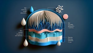
  4. Barrier Function of the Stratum Corneum -

    The "brick-and-mortar" model describes corneocytes as bricks and intercellular lipids as mortar, forming a waterproof barrier that prevents transepidermal water loss. Studies from the National Institute of Arthritis and Musculoskeletal and Skin Diseases emphasize ceramides' role in maintaining this barrier, a high-yield topic on any integumentary system exam. Sketching this model helps you visualize and ace those integumentary system quizzes.

  5. Phases of Wound Healing -

    Wound repair occurs in four phases - Hemostasis, Inflammation, Proliferation, and Remodeling - often recalled by the acronym "H I P R." The Journal of Dermatological Science highlights growth factors like PDGF in the proliferation phase, a concept that frequently appears in integumentary quiz questions. Create flashcards for each phase to boost confidence and accuracy on your next practice test.

Powered by: Quiz Maker