Fetal membranes + gastrulation

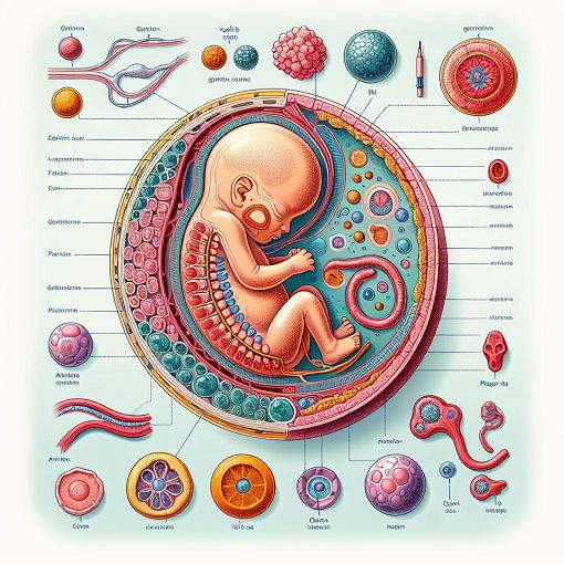
A detailed illustration of human fetal membranes and the gastrulation process, showcasing structures like the yolk sac, amnion, and placenta, with colorful annotations and labels

Understanding Fetal Membranes and Gastrulation

This quiz is designed to test your knowledge of fetal membranes and the process of gastrulation. It covers key concepts, functions, and structures involved in early embryonic development.

  • Assess your understanding of fetal membranes.
  • Explore the role of the placenta in fetal development.
  • Engage with questions on the process of gastrulation and germ layer formation.
15 Questions4 MinutesCreated by StudyingBiology521
Fetal membranes:
- Most of them are derived from the zygote
- Allantois is involved in early hematopoiesis
- Chorion is derived from the trophoblasts
- Are involved in gas exchange
- Amnion is derived from the epiblast
- Mediate in removal of metabolic product of embryo /fetus
- In general they do not form the embryo (with small expections)
- All of them are derived from zygote
- Chorion is derived from the embryonic
- Amnion is derived from the hypoblast
- Amnion is derived from the trophoblast
Chorion, find true
- Is included in formation of amnion
- Is part of yolk sac
- Is formed by cytotrophoblast
- Is included in formation of chorionc villi
- Is formed by synctiotrophoblast
- Secondary villus has a core made of ectoderm
- Is formed only by synctiotrophoblast
- Is formed by cyto- and syncytiotrophoblast
- Secondary villus has a core made by mesenchyme
- Is divided into smooth (leave) and villous (frondosum)
- Only branch, not stem villi are formed from chorion
- Is part of chorionic sac
Allantois, find true
- During the second month, the extraembryonic part of allantois degenerates
- Is a small diverticulum of the caudal part of yolk sac
- Together with yolk sac is included in formation of gut
- Gives rise to oogonia and spermatpgonia
- Differentiates in the ureter
- Can produce urine in prenatal life
- Its blood vessels persist in umbilical cord
- Is involved in early hematopeoiesis
To functions of placenta belong/belongs:
- Transfer gases
- Transfer carbon dioxide
- IgA transport
- Synthesis of LH
- Transfer oxygen
- Synthesis of immunoglobulins
- IgG transport
- Nutritional substance support
Functions of fetal membranes, find true:
- Yolk sac – its derivatives is an epithelium of the respiratory tract
- Yolk sac – source of primordial germ cells
- Allantois – formation of gut
- Amnion – early hematopoiesis
- Amnion – source of primordial germ cells
- Smooth chorion – fetal part of placenta
- Yolk sac – early hematopoiesis
Find the right matches:
- Yolk sac – its derivates is an epithelium of respiratory tract
- Amnion – early hematopoiesis
- Smooth chorion – fetal part of placenta
- Yolk sac – source of primordial germ cells
- Amnion – environment for embryo/fetus development
- Yolk sac – early hematopoiesis
- Allantois – formation of gut
Amnion – find true:
- Its amniotic fluid sac is filled with amniotic fluid
- Up to week 20 amniotic fluid is similar to fetal serum
- Fetus do not participate in production of fluid
- Is filled with about 2,5l of fluid
Yolk sac:
- Is derived from hypoblast
- Is involved in gut formation
- Primordial germ cells appear among the endodermal cells of its wall
- Primitive Yolk sac is fetal part of placenta
- Is responsible for nourishment of the embryo during first 4 month of development
- Is derived from epiblast
- Is responsible for the nourishment of the embryo during the early stages of development
- Is small and devoid of yolk
- Is lined by endoderm and mesenchyme
- Accumulates yolk
Yolk sac, find true:
- During the fourth week, the endoderm of yolk sac is incorporated into the ember as the primordial gut - The epithelium of the digestive tract, trachea, bronchi, lungs
- It gives rise to primordial germ cells
- Derived from hypoblast
- Is lined by endoderm and mesenchyme
- Primordial germ cells appear among the endodermal cells of its wall
- Involved in gut-formation
- In human, does not accumulate yolk
- Is involved in hematopoiesis
- Is involved in transfer of nutrients to embryo during 2nd and 3rd week
Gastrulation:
- Cells of epiblast form ectoderm
- Includes migration of cells
- Takes place in third week of embryo`s development
- Is process that establishes al three germ layers in embryo
- As its result, ectoderm, mesoderm and endoderm is being developed
- Begins with formation of primitive steak
- Is formation of epiblast layer and hypoblast layer
- Takes place in 6th week of embryos development
- Takes place in the first week of embryo`s development
- Ectoderm is formed during this process (not only)
- Takes place in 3rd week of embryos development
Gastrulation:
- Begins with formation of primitive steak
- Is process that establishes all three germ layers in embryo
- Take place in third week of embryo`s development
- Takes place in first week of development
- Takes place in 6th week of development
- Ectoderm is only formed during this process
- Cells of epiblast form ectoderm
- Includes migration cells
Mesoderm in gastrulation:
- Lateral divides into dermatome and myotom
- Intraembryonic is located between ectoderm and endoderm
- Lateral divides into: dermatome, myotom and sclerotom
- Pariaxial into dermatome, myotom and sclerotom
Development of three germ layers:
- Is called gastrulation
- Endoderm derives from extraembryonic mesoderm
- Intraembryonic mesoderm derives from epiblast
- Cells of epiblast migrate towards primitive streak
- Development of endoderm is first than invagination into epiblast layer
Somites:
- Establish the segmental organisation of the body.
- Derive from somitomers of lateral mesoderm
- Derive from somitomers of paraxial mesoderm
- Derive from endoderm
- At the end of gastrulation there are 17-20 of somites
- At the end of gastrulation there are ca. 50 of somites
Mesoderm:
- Cornea develops from intermediate mesoderm
- Intermediate divides into: dermatome, myotom and sclerotom
- Intraembryonic is located between ectoderm and endoderm.
- Lateral divides into dermatome and myotome
- Lateral divides into: dermatome, myotome and sclerotome (paraxial
- Mesenchyme develops from sclerotom
- Pariaxial divides into: dermatome, myotome and sclerotome.
{"name":"Fetal membranes + gastrulation", "url":"https://www.quiz-maker.com/QPREVIEW","txt":"This quiz is designed to test your knowledge of fetal membranes and the process of gastrulation. It covers key concepts, functions, and structures involved in early embryonic development.Assess your understanding of fetal membranes.Explore the role of the placenta in fetal development.Engage with questions on the process of gastrulation and germ layer formation.","img":"https://cdn.poll-maker.com/104-5107565/img-ebqfrgcxkcqymankcljeemyo.jpg"}
Powered by: Quiz Maker