Part 3 Oral path Identify

A detailed illustration of various oral cysts and dental anomalies with labels, set against a medical background

Oral Pathology Identification Quiz

Test your knowledge of oral pathology with this comprehensive quiz! Designed for dental students and professionals, this quiz explores various conditions, cysts, and anomalies encountered in oral pathology.

Featuring 20 questions, including multiple choice and descriptive formats, you'll cover key topics such as:

  • Cysts and their histological features
  • Dental anomalies and deformities
  • Common oral lesions

20 Questions5 MinutesCreated by IdentifyingLeaf732
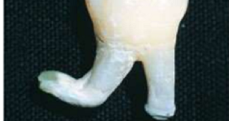
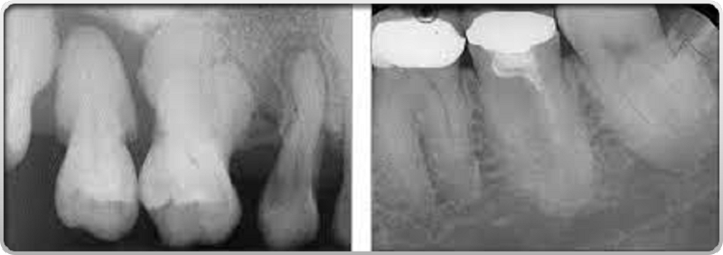
The deformity seen here with a bend in a root apices is characteristic of
Dilaceration
Gemination
Fusion
Concrescence
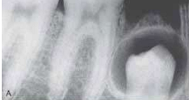
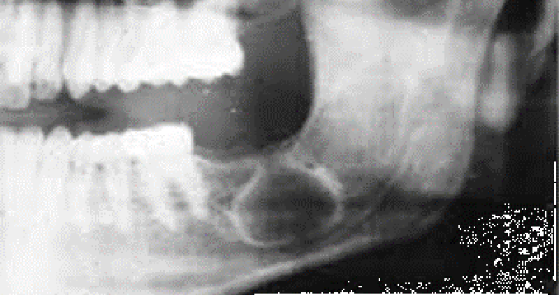
This unilocular radiolucency around the crown of an unerupted second premolar is most likely a
Normal developmental sac
Dentigerous cyst
Primordial cyst
Lateral periodontal cyst
Most common oral cyst
Dentigerous cyst
Eruption cyst
Primordial cyst
Radicular cyst
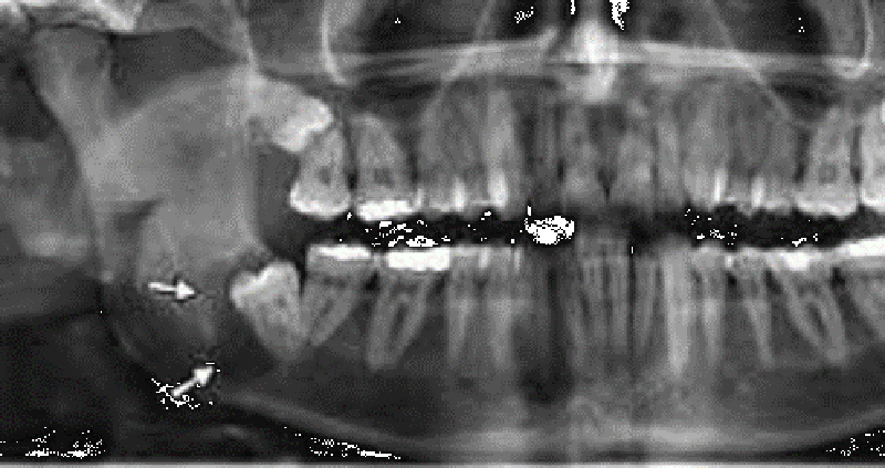
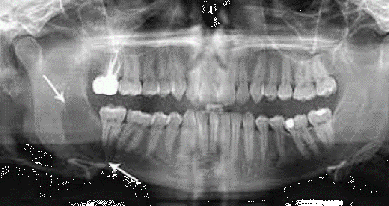
Most commonly occurs around the crown of an unerupted or impacted third molar
Eruption cyst
Dentigerous cyst
Radicular cyst
Primordial cyst
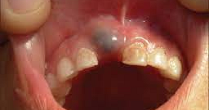
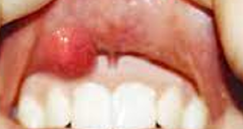
Found in the soft tissue around the crown of an erupting tooth
Eruption cyst
Dentigerous cyst
Radicular cyst
Primordial cyst
Most commonly in place of a third molar
Primordial cyst
Eruption cyst
Dentigerous cyst
Radicular cyst
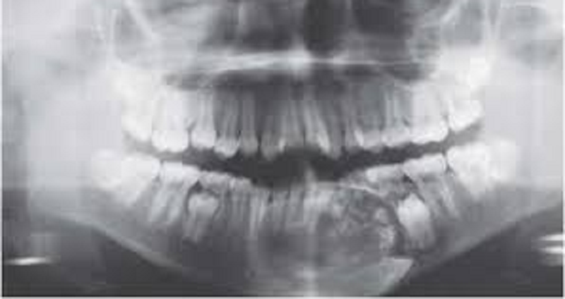
Has a unique histologic appearance and high recurrence rate
Primordial cyst
Keratocyst (OKC)
Calcifying Odontogenic cyst (COC)
Radicular cyst
Has a characteristic feature called ghost cells
Primordial cyst
Keratocyst ( OKC)
Calcifying Odontogenic cyst (COC)
Radicular cyst
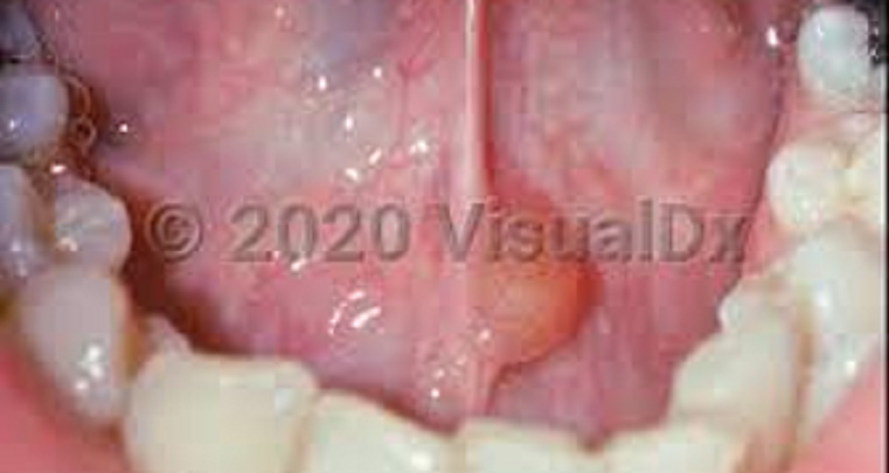
An expansion or swelling in the mucobuccal fold in the area of the maxillary canine and the floor of the nose
Nasolabial cyst
Lymphoepithelial cyst
Dermoid cyst
Thyroglossal cyst
Appears to arise from epithelium trapped in a lymph node during development
Nasolabial cyst
Lymphoepithelial cyst
Dermoid cyst
Thyroglossal cyst
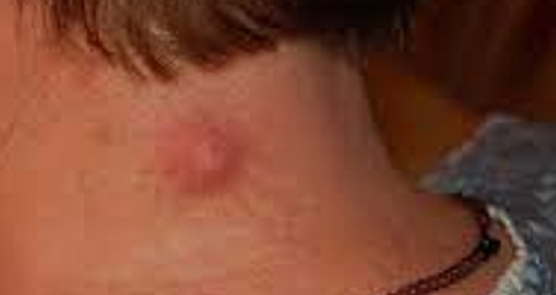
A raised nodule on the skin of the face or neck
Nasolabial cyst
Lymphoepithelial cyst
Epidermal cyst
Dermoid cyst
Located between the roots of the maxillary lateral incisor and cuspid
Globulomaxillary cyst
Dermoid cyst
Epidermal cyst
Thyroglossal cyst
Tooth #7 is an example of
Tooth #8 is an example of
Appears as two crowns joined together by a notched incisal area
Excessive cementum on the roots of the teeth
Cementum
Hypocementosis
Hypercementosis
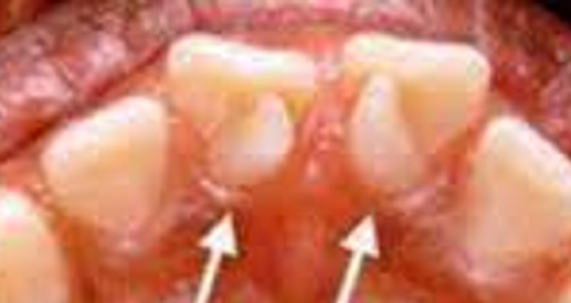
An accessory cusp located in the cingulum area of a maxillary or mandibular permanent incisor
Cingulum
Ligual cusp
Talon cusp
The teeth have elongated pulp chambers and short roots
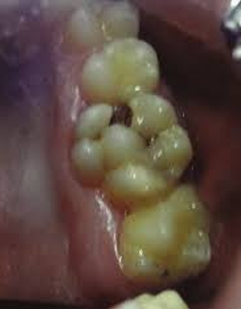
An accessory enamel cusp found on the occlusal tooth surface
Dens Invaginatus
Dens Evaginatus
Often accompanies a periapical lesion
{"name":"Part 3 Oral path Identify", "url":"https://www.quiz-maker.com/QPREVIEW","txt":"Test your knowledge of oral pathology with this comprehensive quiz! Designed for dental students and professionals, this quiz explores various conditions, cysts, and anomalies encountered in oral pathology.Featuring 20 questions, including multiple choice and descriptive formats, you'll cover key topics such as: Cysts and their histological features Dental anomalies and deformities Common oral lesions","img":"https:/images/course2.png"}
Powered by: Quiz Maker