EMT Anatomy and Physiology Quiz: Test Core Body Systems
Quick, free EMT anatomy quiz with instant results and feedback.
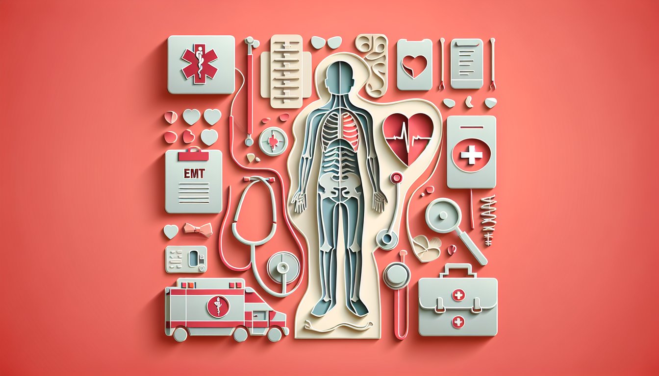
This quiz helps you review EMT anatomy and physiology you need in the field, with questions on body systems and function. Get quick results and pinpoint topics to study next, like terminology, tissues, and shock. For extra practice, try our anatomical terminology quiz, dive into the tissues practice test, or check your understanding with the types of shock quiz before moving on.
Study Outcomes
- Understand Skeletal and Muscular Frameworks -
Identify and recall major bones, joints, and muscle groups critical for patient movement and stabilization within emt anatomy.
- Analyze Organ System Functions -
Differentiate the roles of respiratory, cardiovascular, and nervous systems through an emt anatomy and physiology lens to better assess patient conditions.
- Interpret Medical Terminology -
Decipher common directional terms, body planes, and medical vocabularies featured in this anatomy quiz for emt to communicate effectively with healthcare teams.
- Apply EMT Medical Anatomy to Patient Assessment -
Use knowledge of anatomical landmarks and structures to perform physical examinations and recognize injury patterns in real-world emergencies.
- Reinforce Physiology Concepts -
Solidify your grasp of organ and tissue functions under normal and stress conditions by tackling questions in the emt physiology quiz.
- Enhance Confidence in Emergency Scenarios -
Build assurance in your emt anatomy knowledge and practical skills, empowering you to respond swiftly and accurately during calls.
Cheat Sheet
- Skeletal System Landmarks -
Review the 206 bones of the body, focusing on key EMT anatomy landmarks like the skull, spine, sternum, and pelvis as outlined in Gray's Anatomy and university anatomy courses. Use the mnemonic "Some People Study Science Periodically" to recall Skull, Spine, Sternum, Scapula, Pelvis. Mastering these landmarks helps you spot fractures and apply proper splinting and immobilization techniques in the field.
- Cardiovascular Physiology Basics -
Understand how the heart works by memorizing the cardiac output equation (CO = HR × SV), where CO is cardiac output, HR is heart rate, and SV is stroke volume, as described by the American Heart Association. Knowing emt physiology concepts like preload, afterload, and contractility lets you assess shock and use interventions such as IV fluids or vasopressors appropriately. Visualize the heart's four chambers and flow direction - right atrium → right ventricle → lungs → left atrium → left ventricle - for swift decision-making in emergencies.
- Respiratory Anatomy and Gas Exchange -
Review airway structures from the nasal cavity to the alveoli, using the memory phrase "Never Let Monkeys Eat Bananas" (Nose, Larynx, Mouth, Epiglottis, Bronchi). Emphasize the alveolar-capillary interface where O₂ and CO₂ swap across a one-cell-thick membrane, a concept highlighted in EMT textbooks and university physiology courses. Efficient airway management and ventilatory support depend on pinpointing anatomical obstructions and understanding gas diffusion principles.
- Muscular System and Movement -
Differentiate skeletal, smooth, and cardiac muscle, focusing on the major skeletal muscle groups - biceps, triceps, quadriceps, and hamstrings - as taught in reputable EMT anatomy curricula. Use the phrase "Biceps Bend, Triceps Travel" to remember that biceps flex the elbow while triceps extend it. Familiarity with muscle origin, insertion, and action supports proper patient handling, trauma assessment, and movement injury prevention.
- Nervous System Divisions -
Break down the central and peripheral nervous systems, then subdivide the autonomic system into sympathetic ("fight or flight") and parasympathetic ("rest and digest") branches, as described by the National Institutes of Health. Remember "S for Stress, P for Peace" to recall their opposing effects on heart rate, pupil size, and digestion. Quick recognition of neurological signs and autonomic responses boosts your assessment accuracy for head injuries, stroke, and shock.