Unlock hundreds more features
Save your Quiz to the Dashboard
View and Export Results
Use AI to Create Quizzes and Analyse Results

Sign inSign in with Facebook
Sign inSign in with Google

Take the Eye Anatomy Quiz - Label Parts of the Eye Now!

Think you can ace this eyeball diagram quiz? Dive in and master the parts of the eye now!

Difficulty: Moderate
2-5mins
Learning OutcomesCheat Sheet
Paper art illustration for eye anatomy quiz on teal background

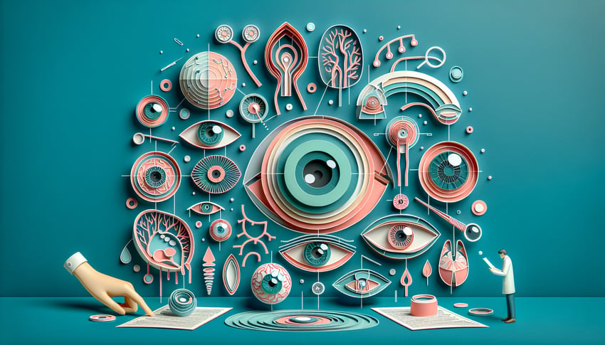
This eye anatomy quiz helps you practice labeling every part of the eye on a diagram, from cornea and iris to retina and optic nerve. Use it to check gaps before a lab or exam and lock in key terms through quick, repeatable rounds.

Which part of the eye is the transparent front covering that helps focus light?
Retina
Iris
Cornea
Sclera
The cornea is the clear, dome-shaped surface covering the front of the eye and is responsible for most of the eye's refractive power. It works together with the lens to focus light precisely on the retina. Unlike the sclera, the cornea is transparent to allow light entry. .
Which structure controls the amount of light entering the eye by adjusting the size of the pupil?
Optic nerve
Cornea
Lens
Iris
The iris is the colored ring of muscle fibers that contract or dilate to change the pupil's diameter, regulating light entry. The lens focuses light but does not adjust aperture size. The optic nerve transmits signals to the brain rather than controlling light. Explore iris function.
What is the name of the opening in the center of the iris?
Pupil
Retina
Macula
Lens
The pupil is the central aperture within the iris through which light passes into the eye. Its size is regulated by iris muscles. The lens sits behind the pupil and focuses light on the retina. .
Which part of the eye contains photoreceptor cells that detect light?
Vitreous humor
Retina
Choroid
Cornea
The retina is the innermost layer of the eye, containing rods and cones that convert light into electrical signals. The choroid provides blood supply but does not detect light. The cornea is an external refractive surface, and the vitreous humor is a gel filling the eye. .
Which structure changes shape to focus light onto the retina?
Iris
Lens
Optic disc
Cornea
The lens is a flexible, transparent structure that adjusts its curvature via the ciliary muscles to focus light onto the retina. The cornea provides fixed refractive power. The iris controls light entry but does not focus. .
What is the gel-like substance filling the space between the lens and retina?
Vitreous humor
Sclera
Aqueous humor
Choroid
The vitreous humor is a clear gel that fills the space behind the lens and supports the retina. The aqueous humor is a fluid in the anterior chamber between the cornea and lens. The sclera is the white outer wall, and the choroid is a vascular layer. .
Which layer of the eye provides nutrients to the retina and contains blood vessels?
Cornea
Choroid
Retina
Sclera
The choroid is the vascular layer between the sclera and retina, supplying oxygen and nutrients to the outer retina. The sclera is primarily protective, and the cornea is avascular. The retina receives its inner blood supply from different vessels. .
The blind spot, where the optic nerve exits the retina, is known as the:
Fovea
Lamina cribrosa
Macula
Optic disc
The optic disc is the point on the retina where ganglion cell axons exit as the optic nerve, creating a physiologic blind spot. The macula and fovea are areas of high acuity vision, and the lamina cribrosa is part of the sclera where fibers pass through. .
What is the central pit in the macula responsible for high-acuity vision?
Optic disk
Ora serrata
Ciliary body
Fovea centralis
The fovea centralis is a small depression in the macula densely packed with cones, providing the highest visual acuity and color discrimination. The optic disk is the blind spot, the ciliary body controls lens shape, and the ora serrata is the retinal boundary. .
Which structure consists of smooth muscle fibers that attach to the lens to facilitate accommodation?
Choroid
Ciliary body
Trabecular meshwork
Zonular fibers
The ciliary body contains smooth muscle that alters lens tension for focusing. Zonular fibers connect the ciliary body to the lens but are not muscle. The trabecular meshwork drains aqueous humor, and the choroid is a vascular layer. Ciliary body function.
The junction between the cornea and sclera is known as what?
Limbus
Lacon
Canal of Schlemm
Palpebral fissure
The limbus is the transitional border where the clear cornea meets the opaque sclera. The Canal of Schlemm drains aqueous humor, the palpebral fissure is the eyelid opening, and 'Lacon' is not a recognized anatomical term. .
Which structure drains aqueous humor from the anterior chamber into the canal of Schlemm?
Trabecular meshwork
Vitreous base
Schlemm's canal
Uveal tract
The trabecular meshwork is a spongy tissue located at the angle of the anterior chamber that filters aqueous humor into Schlemm's canal. Schlemm's canal is the collecting channel but not the initial filter. The uveal tract covers the iris and ciliary body, and the vitreous base supports the retina. Trabecular meshwork function.
Which photoreceptor cells are responsible for color vision and are concentrated in the fovea?
Rods
Bipolar cells
Ganglion cells
Cones
Cones are photoreceptors that mediate color vision and are densely packed in the fovea for high-resolution sight. Rods function in low-light conditions but do not detect color. Ganglion and bipolar cells process signals but are not photoreceptors. .
The blood supply to the inner retina is primarily delivered by which vessels?
Central retinal artery
Choroidal vessels
Ciliary arteries
Vortex veins
The central retinal artery enters the eye via the optic nerve to supply the inner retinal layers with oxygenated blood. Choroidal vessels nourish the outer retina, ciliary arteries supply the uvea, and vortex veins drain choroidal blood. .
0
{"name":"Which part of the eye is the transparent front covering that helps focus light?", "url":"https://www.quiz-maker.com/QPREVIEW","txt":"Which part of the eye is the transparent front covering that helps focus light?, Which structure controls the amount of light entering the eye by adjusting the size of the pupil?, What is the name of the opening in the center of the iris?","img":"https://www.quiz-maker.com/3012/images/ogquiz.png"}

Study Outcomes

  1. Identify Key Eye Structures -

    Learn to recognize and name the major parts of the eye, including the cornea, iris, retina, and lens, through targeted labeling challenges.

  2. Label an Eyeball Diagram Accurately -

    Practice pinpointing each component on an eyeball diagram to reinforce spatial understanding of ocular anatomy.

  3. Describe Functional Roles -

    Explain the primary functions of each eye structure, such as light refraction by the cornea and light detection by the retina.

  4. Differentiate Anatomical Regions -

    Distinguish between the anterior and posterior segments of the eye to deepen comprehension of internal and external eye anatomy.

  5. Apply Anatomical Terminology -

    Use proper ophthalmic vocabulary when discussing eye parts, ensuring clear communication in academic and clinical contexts.

  6. Assess and Improve Knowledge Gaps -

    Identify areas of weakness through quiz results and target specific eye anatomy topics for further review.

Cheat Sheet

  1. Cornea: The Transparent Window -

    The cornea, referenced by the American Academy of Ophthalmology, is the eye's clear, dome-shaped front layer that refracts about two-thirds of incoming light. Remember it in the eye anatomy quiz as the "Clear Cover" using the mnemonic "Cornea Clears" to recall its protective and focusing role. Practicing with an eyeball diagram quiz will help you pinpoint its position and curvature quickly.

  2. Iris & Pupil: Light Regulators -

    The iris, often featured in parts of the eye quiz questions, is the colored ring that adjusts pupil size to control light entry, much like a camera shutter. Use the mnemonic "Iris Illuminates" to link its color and function, and quiz yourself by labeling the iris and pupil in a parts of an eye quiz. Try alternating light conditions in an eye quiz to reinforce how the pupil dilates and constricts under different stimuli.

  3. Lens & Accommodation: Focusing Mechanism -

    Located behind the iris, the lens fine-tunes focus by changing shape through ciliary muscle contraction, a principle highlighted in university-level anatomy resources. Recall "Accommodate to Communicate" to remember accommodation when studying for your eye anatomy quiz or eyeball diagram quiz. Diagram drills labeling the lens in near and distance focus scenarios can cement the concept quickly.

  4. Retina & Photoreceptors: Image Capture -

    The retina, detailed in texts from the National Eye Institute, contains rods for low-light vision and cones for color and detail, with the fovea being the cone-rich central pit. Use "Rods Roam, Cones Close" to distinguish peripheral versus central roles when tackling a parts of the eye quiz. Running through mock questions on rod/cone distribution in an eye anatomy quiz strengthens retention.

  5. Optic Nerve & Pathway: Signal Transmission -

    The optic nerve carries visual signals from the retina to the brain and crosses at the optic chiasm, a fact emphasized in peer-reviewed neuro-ophthalmology studies. Remember "X-chiasm, X-cite" to link the crossing fibers with signal excitement when practicing an eyeball diagram quiz. Quick-draw exercises mapping the optic pathways on blank eye diagrams aid speedy recall during your eye quiz.

Powered by: Quiz Maker