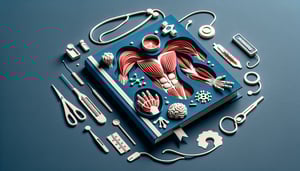
Muscles of the Arm Quiz: Identify and Label Each Muscle
Quick, free arm muscles labeling quiz with instant feedback.
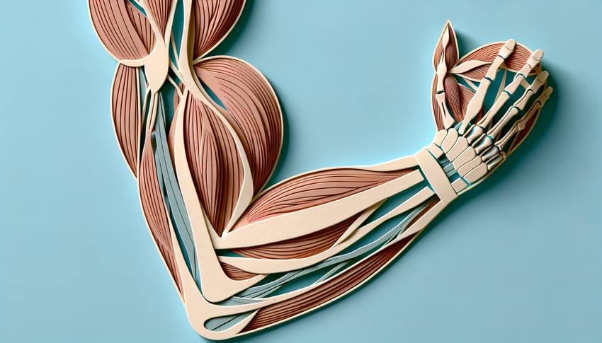
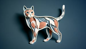
This quiz helps you label and locate the muscles of the arm fast. Build recall on biceps, triceps, and forearm groups with clear images and instant results. For more focused practice, try the forearm muscles quiz , review the upper arm anatomy quiz , or challenge yourself to identify the highlighted muscle.
Study Outcomes
- Identify Major Arm Muscles -
Pinpoint and label key muscles such as the biceps brachii, triceps brachii, and brachialis on an arm diagram.
- Differentiate Forearm Muscle Groups -
Distinguish between the flexor and extensor muscle groups of the forearm and place them correctly in your labels.
- Apply Anatomical Terms in Labeling -
Use precise anatomical terminology for muscle origins, insertions, and actions to enhance the accuracy of your arm muscle labeling.
- Recall Functions of Arm and Forearm Muscles -
Memorize and state the primary actions of each muscle, including flexion, extension, pronation, and supination.
- Analyze Muscle Coordination in Arm Movements -
Understand how different arm and forearm muscles work together during common movements like lifting, pushing, and pulling.
- Evaluate Your Labeling Accuracy -
Test your knowledge with the muscles of the arm quiz to receive immediate feedback and a score that highlights your mastery and areas for improvement.
Cheat Sheet
- Mastering the Biceps Brachii -
The biceps brachii has two heads (long and short) that originate on the scapula and insert on the radial tuberosity, making it the primary elbow flexor. Remember "SLiDER" (Short head - Coracoid, Long head - Supraglenoid) to ace your muscles of the arm labeling. Its role in supination is tested often in any muscles of the arm quiz, so visualize the twisting motion of the forearm.
- Understanding the Triceps Brachii -
The triceps brachii extends the elbow and consists of long, lateral, and medial heads that converge on the olecranon process. A simple mnemonic "3 Heads => 1 Tee" helps you recall the three origins and single insertion point. In arm muscle labeling drills, look for the posterior compartment bulk and olecranon landmarks.
- Spotlighting Brachialis and Brachioradialis -
The brachialis lies deep to the biceps and is a pure flexor of the elbow, while the brachioradialis spans from the lateral supracondylar ridge to the styloid process of the radius. Use "Beer Raising" to remember brachioradialis action when lifting a glass - perfect for forearm muscles quiz practice. Labeling these correctly is key to distinguishing deep versus superficial flexors.
- Deep Flexors & Pronators of the Forearm -
In the anterior compartment, muscles like pronator teres, flexor carpi radialis, and palmaris longus work in concert for wrist flexion and pronation. The phrase "Pass, Fail, Pass" (Palmaris, Flexor carpi radialis, Pronator teres) helps when tackling arm muscle labeling tasks. These deep flexors often appear in forearm muscles quiz questions on tendinous arches.
- Key Extensors in the Posterior Compartment -
Extensor carpi radialis longus/brevis, extensor digitorum, and extensor carpi ulnaris share the common extensor tendon at the lateral epicondyle. Recall "Lynx, Dogs, Unfortunately" (Longus, Digitorum, Ulnaris) to streamline your muscles of the arm quiz answers. Spotting the muscle bellies along the dorsal forearm is crucial for accurate arm labeling.