Identify the Highlighted Muscle: Quick Anatomy Quiz
Quick muscle IQ quiz with images; check the action of the highlighted muscle. Instant results.
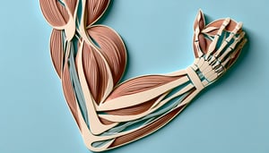
This quick quiz helps you identify the highlighted muscle in each image and recall its action and attachments. Use it to spot weak areas before a test, then build confidence with focused practice in our arm muscles labeling quiz, leg muscle labeling quiz, and abdominal muscles quiz .
Study Outcomes
- Identify the Highlighted Muscle -
Accurately identify the highlighted muscle in anatomical images by selecting which muscle is highlighted based on its shape, location, and attachments.
- Determine Muscle Function -
Identify the action of the highlighted muscle and explain how it contributes to movements such as facial expressions or limb motions.
- Analyze Muscular Attachments -
Answer questions like which of the following muscles inserts on the highlighted structure and locate key origin and insertion points for muscles such as the Masseter.
- Differentiate Similar Muscles -
Distinguish between muscles with overlapping regions to confidently solve which structure is highlighted masseter versus adjacent muscles.
- Apply Anatomical Knowledge -
Use your understanding of muscle anatomy to interpret detailed images and strengthen your ability to identify the highlighted muscle in various contexts.
- Evaluate Your Mastery -
Assess your proficiency through scored quiz feedback and track areas for improvement in identifying muscle names and functions.
Cheat Sheet
- Tracing Origin and Insertion -
When you identify the highlighted muscle, start by locating its bony landmarks - origins often hide beneath other tissues. For example, the zygomatic arch and mandible are key to pinpointing the masseter (University of Michigan Anatomy). Use the mnemonic "FIDO" (From Insertion, to Origin) to reverse-trace attachments when stumped.
- Recognizing Fiber Direction -
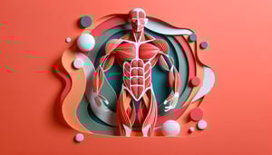
Muscle fiber orientation (parallel, pennate, convergent) is a reliable clue to determine which muscle is highlighted. The external oblique's inferior fibers run "hands-in-pockets" while the rectus abdominis fibers run straight up and down (Gray's Anatomy). Noting fiber angle helps you identify the action of the highlighted muscle quickly.
- Surface Landmark Palpation -
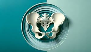
Palpate bony landmarks like the anterior superior iliac spine or the lateral epicondyle to find nearby muscle bellies in cadaver labs. When asked "which muscle is highlighted," use surface anatomy guides from the American Association of Clinical Anatomists. This hands-on approach cements your spatial understanding.
- Innervation Clues -
Knowing which nerve innervates a muscle narrows the field - for instance, the facial nerve's masseteric branch signals the masseter, while the radial nerve targets the triceps brachii (Netter's Atlas). If you're quizzed on "identify the action of the highlighted muscle," pair the nerve with its primary movement. Nerve roots act as a fail-safe when structure alone isn't enough.
- Functional Testing Techniques -
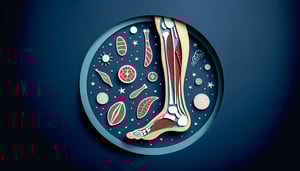
Use manual muscle testing to verify which muscle is highlighted: resist plantarflexion to isolate the gastrocnemius versus the soleus when the knee is extended or flexed (American Physical Therapy Association). Asking "which of the following muscles inserts on the highlighted structure" becomes easier once you link movement to muscular pull. Practice active motion exams to sharpen your clinical identification skills.