Anterior Thigh Muscles Quiz: Label the Major Muscles
Quick thigh muscles quiz to test your anatomy skills. Instant results.
This quiz helps you label the anterior thigh muscles and see which names you know cold. You will practice the major muscles on the front of the thigh and get instant feedback. Use it before lab or a test, then deepen your review with label the leg muscles and the leg muscles anatomy quiz.
Study Outcomes
- Identify key anterior of thigh muscles -
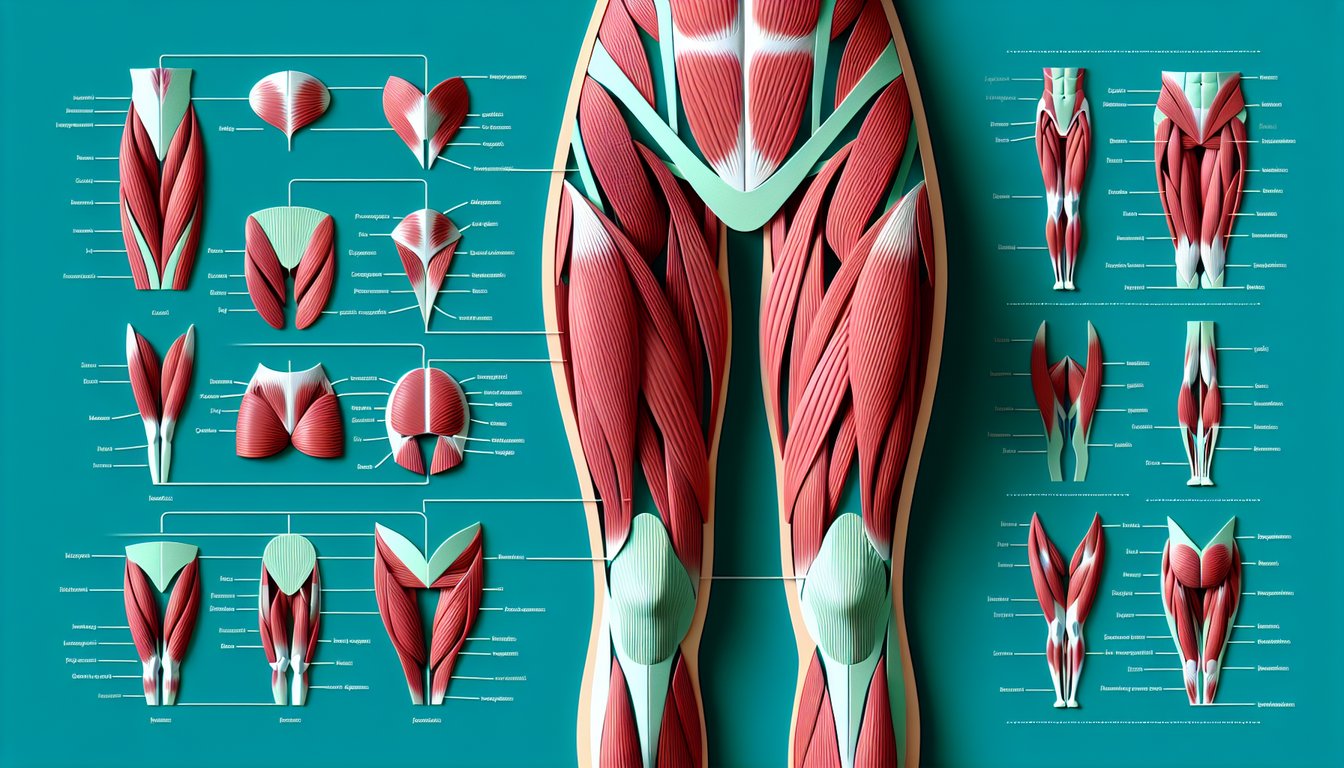
Pinpoint and correctly label the major front thigh muscles such as the quadriceps group and sartorius in anatomy diagrams.
- Describe anterior thigh anatomy landmarks -
Outline the origins, insertions, and anatomical boundaries that define the anterior compartment of the thigh.
- Explain muscle functions -
Detail the primary roles of each anterior thigh muscle in movements like knee extension and hip flexion.
- Apply labeling skills in a quiz format -
Use interactive questions to reinforce accurate muscle identification and cement your knowledge under timed conditions.
- Assess your mastery of front thigh muscles names -
Review instant feedback and quiz scores to determine which areas of anterior thigh anatomy require further study.
Cheat Sheet
- Quadriceps Femoris Composition -
The quadriceps femoris group is the powerhouse of the anterior of thigh, comprising rectus femoris, vastus lateralis, vastus medialis, and vastus intermedius. A friendly mnemonic "Really Very Vastly Internally" helps you correctly label the anterior thigh muscles in order from superficial to deep. These fibers unite into the quadriceps tendon that inserts on the patella, enabling powerful knee extension during activities like jumping and squatting.
- Sartorius: The Longest Muscle -
In the anterior of thigh, the sartorius is the longest muscle, running from the anterior superior iliac spine to the medial tibia. As described in Gray's Anatomy, it flexes, abducts, and externally rotates the hip while assisting knee flexion - picture the "tailor's stitch" pose to lock in this multi-joint action. This biarticular mechanism is essential for movements like crossing your legs and providing dynamic stability.
- Femoral Nerve Innervation -
Anterior thigh muscles draw their innervation from the femoral nerve (roots L2 - L4), which travels under the inguinal ligament into the thigh. Remember the "NAVEL" mnemonic (Nerve, Artery, Vein, Empty space, Lymphatics) to locate the nerve lateral to the femoral artery in the femoral triangle. Testing the patellar reflex offers a quick, clinical way to assess femoral nerve integrity during patient exams.
- Femoral Artery and Blood Supply -
Blood supply to the anterior of thigh stems from the femoral artery as it enters the femoral triangle, with deep branches like the profunda femoris feeding the quadriceps. Use the inguinal ligament as a reliable landmark - the artery lies just beneath it when entering the thigh. Palpating a strong femoral pulse halfway between the ASIS and pubic symphysis confirms adequate perfusion in clinical settings.
- Active Recall & Q - Angle Assessment -
To master anterior thigh anatomy, practice active recall by correctly labeling the anterior thigh muscles on diagrams - an effective memory boost backed by educational research. Familiarity with the Q - angle (about 14° in men, 17° in women) also helps you understand patellar tracking and knee mechanics. Combining mnemonic techniques with quiz-style repetition turns diagrams into memorable study tools for long-term retention.