Cat Muscles Quiz: Label the Brachialis and More
Quick, free cat muscle labeling quiz. Instant results.
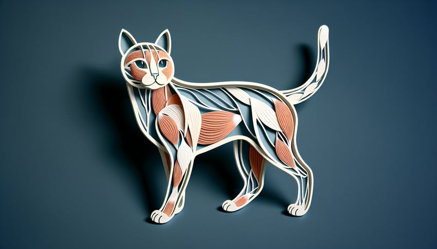
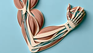
This cat muscles quiz helps you practice labeling feline anatomy and spot the brachialis on diagrams. For more targeted review, try our muscles of the arm quiz, forearm muscles quiz , and upper limb muscles quiz . Get instant feedback and build recall before your next lab or exam.
Study Outcomes
- Identify Key Feline Muscles -
Recognize and name major muscles in cat muscle labeling, including the spinotrapezius and vastus lateralis, to master feline anatomy.
- Locate Muscle Positions -
Pinpoint precise anatomical locations of muscles on an interactive cat anatomy muscles labeled diagram to enhance spatial understanding.
- Differentiate Muscle Functions -
Explain the roles of primary cat muscles during movement and support, reinforcing concepts from the cat muscle anatomy quiz.
- Apply Anatomical Terminology -
Use correct veterinary anatomy terms when discussing muscle groups in a cat dissection quiz to communicate clearly and professionally.
- Evaluate Your Knowledge -
Assess your proficiency with the cat muscles quiz through immediate feedback, identifying strengths and areas for further study.
Cheat Sheet
- Identifying Superficial Back Muscles -
In cat muscle labeling, the spinotrapezius lies caudal to the acromiotrapezius, with fibers running obliquely toward the spine. The acromiotrapezius, positioned between the scapulae, has transverse fibers that help you differentiate it during dissection (University of Wisconsin Dissection Guide). Mastering their orientation is key to acing your cat muscles quiz!
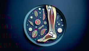
- Distinguishing Hindlimb Extensors -
The quadriceps group, including vastus lateralis and vastus medialis, anchors on the femur and converges on the patella, generating knee extension (Journal of Veterinary Anatomy, 2020). Use the mnemonic "VAM" - Vastus, Articularis, Medialis - to recall their order from lateral to medial. Visualizing this arrangement boosts confidence in your cat muscle anatomy quiz and cat dissection quiz.
- Tracing Muscle Origin and Insertion -
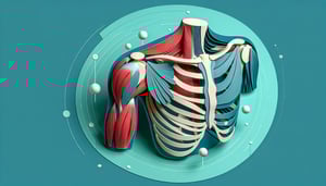
Understanding that the latissimus dorsi originates on the thoracolumbar fascia and inserts on the humerus clarifies its role in forelimb retraction (Smith et al., Comparative Anatomy Journal). By noting bony landmarks, you can predict muscle function before even touching a scalpel. This analytical approach is vital for any cat muscles quiz or cat anatomy muscles labeled exercise.
- Employing Mnemonic Devices -
Use the mnemonic "SALT" (Spinotrapezius, Acromiotrapezius, Levator scapulae ventralis, Trapezius) to sequence dorsal muscles from spine to scapula according to the University of Michigan's Anatomy Lab. Pointing to each muscle while reciting the acronym cements its spatial arrangement in your memory. This fun trick directly enhances performance in cat muscle anatomy quizzes and cat dissection quizzes.
- Mastering Dissection Techniques -
Begin with a clean midline incision and gently reflect the skin to expose underlying musculature without tearing fibers, as recommended by the Association of Veterinary Anatomists. Observing fiber direction (parallel vs. pennate) and texture helps identify each muscle group accurately. Practicing these steps ensures you'll breeze through any cat muscles quiz or cat anatomy muscles labeled challenge.