
DES 2016. Final ( Part 11 )

Pulmonary Medicine Mastery Quiz
Test your knowledge in pulmonary medicine with this comprehensive quiz designed for medical professionals and students alike. With 100 carefully curated questions, you'll explore various critical topics including respiratory diseases, diagnostic methods, and treatment protocols.
Prepare to challenge yourself!
- 100 questions covering key pulmonary topics
- Multiple choice format for easy answering
- Ideal for students and healthcare professionals
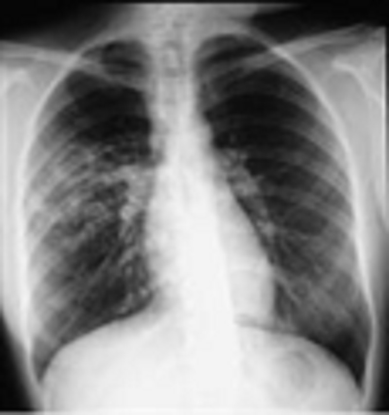
71) Une patiente de 32 ans, vous consulte pour toux et altération de l’état général depuis 2 mois. Elle a perdu du poids 5 Kg en 2 mois. Elle se plaint aussi d’asthénie intense. Elle est mariée un enfant de 3 ans et ne prend aucun traitement d’une pilule oestroprogestative. Le cliché de radio thoracique effectuée en ville . Quelle est l’anomalie sur la radiographie thoracique retrouvez-vous ?
Opacité du lobe moyen
Opacité excavée
Opacité infiltrative
Opacité grelot
Opacité micronodule
72) Vous recevez en consultation une patiente de 60 ans pour une dyspnée expiratoire. Le patient suivi régulier pour une bronchopneumopathie chronique obstructive (BPCO) post tabagique. La dyspnée d’apparition progressive associée à une toux avec expectoration purulente depuis 5 jours qui ne cessent de se majorer. Vous évoquez donc à une exacerbation de bronchopneumopathie chronique obstructive (BPCO) post tabagique. Cliniquement, vous trouvez une légère cyanose des extrémités et des sueurs, la fréquence respiratoire : 26/min, la tension artérielle 140/90 mmHg, la fréquence cardiaque : 90 par min. Quelle est l’étiologie d’origine infectieuse le plus souvent responsable?
Influenza A
Legionella pneumonia
Mycobacterium tuberculosis
Hemophylus influenzae
Bordetella pertussis
73) Une patiente de 62 ans cuisinière, vit seule sans famille, vous suivez une bronchopneumopathie chronique obstructive (BPCO) biomasse stade 3 GOLD, consulte aux urgences pour une dyspnée d’apparition progressive. Vous évoquez donc une exacerbation bronchopneumopathie chronique obstructive (BPCO). Cliniquement, vous trouvez une légère cyanose des extrémités et des sueurs, la fréquence respiratoire : 26/min, la tension artérielle 140/90 mmHg, la fréquence cardiaque : 90 par min. Quelle est l’anomalie sur le cliché de radiographie thoracique trouvé le plus souvent ?
Aplatissement des coupoles diaphragmatique
Diminution diamètre antéro-postérieur
Élargissement silhouette cardiaque
Inspiration insuffisance
Opacité périhilaire
74) Vous recevez aux urgences Mr. S., 35 ans, pour une dyspnée expiratoire. A l’examen clinique, vous trouvez des sibilants diffus. Les constantes sont suivantes : la fréquence cardiaque : 120/min, la tension artérielle : 120/80 mmHg, la fréquence respiratoire : 28/min, la saturation SpO22 : 95% en air ambiant. Le diagnostic un asthme retenu le plus vraisemblablement. Quel est l’examen complémentaire indispensable à l’état stable pour le diagnostic confirmation d’asthme de ce patient ?
Radiographie thoracique
Gaz du sang
TDM thoracique
Explorations fonctionnelles respiratoires
Test allergologique
75) Vous recevez aux urgences Me. H. , 35 ans, pour une dyspnée aigue. Elle est très agitée et est incapable de terminer ses phrases. A l’examen clinique, vous trouvez des sibilants diffus, ainsi qu’une cyanose. Le diagnostic asthme aigu grave retenu le plus vraisemblablement. Le gaz du sang pH : 7,26 PaO2 : 65 mmHg, PaCO2 : 64 mmHg HCO3 29 mmol/l. Quelle anomalie les gaz du sang la plus appropriée ?
Acidose respiratoire
Acidose mixte
Partiellement compensé
Hyperventilation alvéolaire
Shunt vrai anatomique
76) Vous recevez en hospitalisation un chauffeur âgé de 27 ans pour sensation d’étouffement. Poids 109 Kg pour taille 1m62. La tension artérielle: 143/98 mmHg, poules 98 par min, fréquence respiratoire : 21/min et SpO2 96% en état éveil et 84% en dorment avec ronflement. Il n’a pas antécédent particulier ni acromégalie ni hypothyroïdie. Il a deux accidents de voie publique par rapport à la somnolence. Vous pensez à syndrome d’apnée du sommeil. Quel examen permet de faire le diagnostic de syndrome d’apnée du sommeil ?
Electrocardiogramme (ECG)
Enregistrement polysomnographique (PSG)
Electromyogramme (EMG)
Electroencéphalogramme (EEG)
Explorations fonctionnelles respiratoires (EFRs)
77) Vous recevez en hospitalisation un chauffeur âgé de 27 ans pour sensation d’étouffement. Poids 109 Kg pour taille 1m62. La tension artérielle: 143/98 mmHg, poules 98 par min, la fréquence respiratoire : 21/min et la SpO2 96% en état éveil et 84% en dorment avec ronflement. Il n’a pas antécédent particulier ni acromégalie ni hypothyroïdie. Il a deux accidents de voie publique par rapport au somnolence. Vous pensez syndrome d’apnée du sommeil et la confirmation de polysomnographie. Quel paramètre supplémentaires analysé sur un enregistrement polysomnographique ?
Onde S1Q1 pour bloc branche droit
Electromyogramme des muscles du biceps détection hypotonie
Onde lente diffuse par rapport des crises épileptique
Syndrome obstructif réversible sous β-mimétique
Signal des arrêts du flux nasal avec microréveil
78) Vous recevez en hospitalisation un chauffeur âgé de 27 ans pour sensation d’étouffement. Poids 109 Kg pour taille 1m62. La tension artérielle: 143/98 mmHg, poules 98 par min, la fréquence respiratoire : 21/min et la SpO2 96% en état éveil et 84% en dorment avec ronflement. Il n’a pas antécédent particulier ni acromégalie ni hypothyroïdie. Il a deux accidents de voie publique par rapport à la somnolence. Vous pensez à syndrome d’apnée du sommeil et vous proposez le patient pour enregistrement la poly graphie ventilatoire et vous validez et interprétez la polygraphie ventilatoire (PGV). Quelle est la chiffre définie le syndrome d’apnée obstructive du sommeil sévère selon l’index d’apnées-hypopnées (IAH) ?
IAH 20/heure
IAH 30/heure
IAH 40/heure
IAH 50/heure
IAH 60/heure
79) Un patient de 23 ans en hospitalisation au service pneumologie pour fièvre et frisson depuis une semaine. Il est le gardien de la forêt Koh Kong. Depuis 72 heures, il a douleurs abdominales, selles diarrhéiques associées à la lombalgie. L’apparition de dyspnée intense avec cyanose. La 1ère constance : tension artérielle 94/45 mmHg poules 135 par min température axillaire 40°C. Votre hypothèse diagnostic la plus probable parasitose sévère avec localisation pulmonaire. Quel est le parasite peut-on prélever le plus de chance à identifier dans le sang ?
Plasmodium falciparum
Paragonimus westermani
Pneumocystis jerovecii
Toxoplasma gondi
Leishmania donovani
80) Une patiente de 21 ans en consultation pour prurit anal et toux irritante une semaine. Votre hypothèse diagnostic la plus probable parasitose sévère avec localisation pulmonaire. Quel est le parasite peut infecter cette patiente sans augmenter l’éosinophilie dans la numération formule sanguine (NFS) ?
Amibes
Aspergillus
Ankylostomes
Ascaris
Anguillule
81) Vous recevez en consultation une femme noire de 20 ans pour amaigrissement et asthénie. Elle est mariée sans enfant. Le poids est de 43 Kg pour taille 157cm, la tension artérielle est 130/87 mmHg et poules 102 par fréquence respiratoire 25/min SpO2 97% en air ambiant. Le cliché de radiographie thoracique révèle des opacités hilaires et interstitielles bilatérale. Vous pensez à une sarcoïdose. Quelle est l’anomalie biologique trouvée dans l’électrophorèse des protéines sanguine plus probable?
Diminution albuminémie
Augmentation α1-globulines
Diminution α2-globulines
Normalisation β-globulines
Augmentation γ-globulines
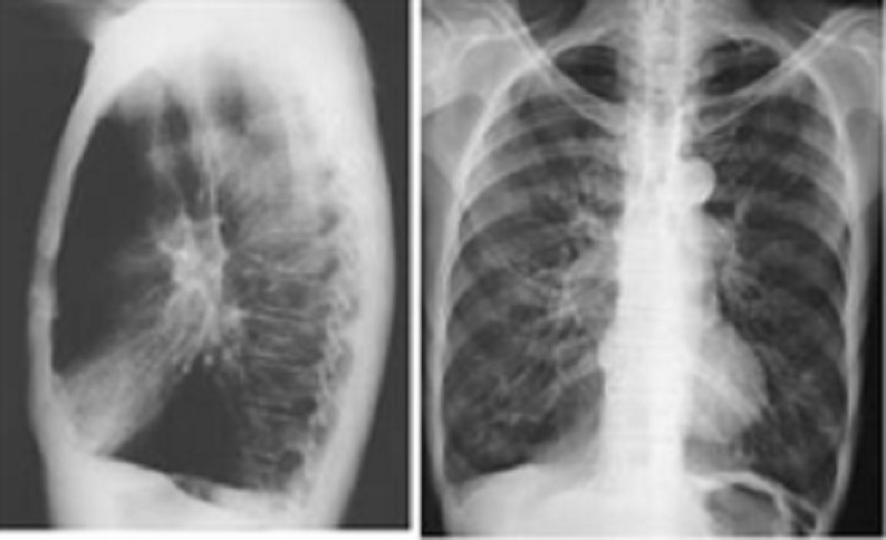
82) Vous recevez en consultation une femme noire de 28 ans pour amaigrissement et asthénie. Elle est mariée sans enfant. Le poids est de 43 Kg pour taille 157cm, la tension artérielle est 130/87 mmHg et poules 102 par fréquence respiratoire 25/min SpO2 97% en air ambiant. Le cliché de radiographie thoracique révèle des opacités hilaires et interstitielles bilatérale. Vous pensez à une sarcoïdose. Quelle est l’anomalie de la tomodensitométrie thoracique la plus adaptée le syndrome de Lofgren stade 1 de la sarcoïdose ?
Adénopathie intrathoracique, bilatérale non compressives
Adénopathie intrathoracique et opacité interstitielle diffus
Opacité interstitielle diffus micronodule ou reticulo-nodulaie
Fibrose pulmonaire avec possibilité de lésions rétractiles
Bronchiectasie localisée et lésion bronchique surinfectée
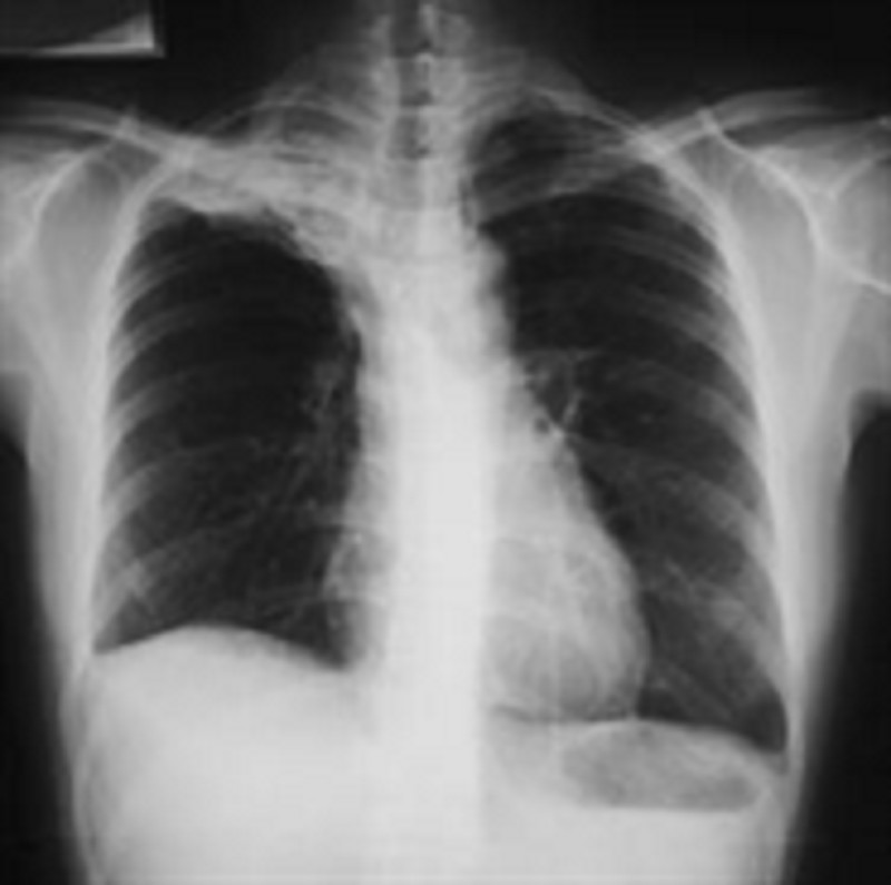
83) Mr. Q., 52 ans vous est adressés par son médecin généraliste. Il se plainte de douleur du membre supérieur droit à type de décharges électriques, qui surviennent le plus souvent la nuit et résistent aux antalgiques usuels (Voltarène et Doliprane). Ces douleurs intéressent la face antéro-interne de l’avant-bras droit ainsi que 2 derniers doigts. Vous observez une amyotrophie de l’éminence hypothénar. Mr. Q. Vous signale également une toux apparue depuis 3 mois accompagnée d’une une grande asthénie et amaigrissement. C’est un patient tabagique à 30 PA. Il vous apporte une radiographie thoracique révèle effectuée en ville. Quel est l’élément retrouvez-vous sur le cliché de radiographie thoracique?
Opacité lobe moyen
Opacité de l’apex
Syndrome bronchique
Lyse osseuse
Adénopathie médiastinale
84) Vous recevez un homme âgé de 57 ans en hospitalisation pour une masse tumorale du médiastin postérieur dans un contexte des douleurs thoraciques apparue récente. Il fume 50 paquet-année. L’examen clinique : conscience normal, la tension artérielle 145/76 mmHg la fréquence respiratoire 24 par min, SpO2 94% en air ambiant. Quel examen paraclinique de référence pour ce patient?
Echographie transthoracique
Radiographie thoracique profile
Tomodensitométrie thoracique
Scintigraphie perfusion
Imagerie résonnant magnétique
85) Vous recevez un homme âgé de 27 ans en hospitalisation pour une masse médiastinale antérieur moyenne dans un contexte d’une oppression thoracique. Il n’a pas antécédent, il ne fume pas. L’examen clinique : conscience normale, la tension artérielle 125/76 mmHg la fréquence respiratoire 24 par min, SpO2 97% en air ambiant. Quel examen physique extra thoracique le plus recherché ?
Palpation air ganglionnaire
Inspection veines jugulaire
Auscultation souffle vasculaire
Inspection hippocratisme digital
Palpation des testicules
86) Vous recevez un homme âgé de 27 ans en hospitalisation pour une masse médiastinale antérieur moyenne dans un contexte d’une oppression thoracique. Il n’a pas antécédent, il ne fume pas. L’examen clinique : conscience normale, la tension artérielle 125/76 mmHg la fréquence respiratoire 24 par min, SpO2 97% en air ambiant, sans altération de l’état général. Quel marqueur tumoral ou biologique le plus dosé?
PSA
ACE
Immunoglobuline
β-2 microglobuline
β hCG
87) A 5 ans, fille est amenée au service d'urgence en Décembre par sa mère, qui se plaint que sa fille semble confuse. La mère signale que sa fille se plaint de maux de tête intermittents puisque les deux d'entre eux déplacés dans le premier étage d'un immeuble ancien il y a 6 mois. La mère a été à la maison avec la fille au cours des dernières 24 heures et la jeune fille semble léthargique et se plaint de douleurs articulaires, des nausées et des maux de tête. Son pouls est de 120 / min, la pression artérielle est 130/85 mm Hg, fréquence respiratoire est de 25 / min, et la saturation en oxygène est de 100% à l'air ambiant. La mère de la fille note également avoir un léger mal de tête qui a commencé hier. Lequel des tests de diagnostic suivants devraient être poursuivis le plus rapidement?
Des gaz du sang artériel
CT scan de la tête
Laryngoscopie directe
ECG
écran de toxicologie
88) Un homme 67 ans, présente � son médecin pour se plaintes de dyspnée � l'effort au cours des 6 derniers mois qui a progressivement de dyspnée au repos. Il nie la toux et une respiration sifflante et n'a pas eu de la fièvre, des sueurs nocturnes, perte de poids involontaire. Il n'a jamais fumé et travaillé comme un constructeur de navires pour> 30 ans.Laquelle des conclusions suivantes sur radiographie thoracique serait confirmé le diagnostic le plus probable?
Infiltrats diffus bilatéraux
Adénopathie hilaire bilatérale
Consolidation du tissu pulmonaire
De masse focale avec bronchogrammes aériens
Des plaques pleurales multiples avec parenchymateuse inégale opacités
89) A 5-month-old child presents to Emergency Department with a reduced conscious level. No history of trauma, but he appears neglected. No physical injury is identified on clinical examination. A CT head is performed. Which one of the following is the most common intracranial finding in non-accidental injury?
Hydrocephalus
Intra-cerebral hemorrhage
Loss of gray-white matter differentiation
Subdural hematoma
Tumor
90) A 7-year-old boy has fallen on an outstretched hand and complains of a painful right elbow. When reviewing the radiographs for evidence of bony injury, which one of the following statements is true?
Posterior fat pad may be a normal finding on flexed lateral view
An anterior fat pad is always abnormal
The line from the anterior cortex of the humerus should pass through the anterior third of the capitellum
The radiocapitellar line should intersect on all views
The radiocapitellar line should not intersect on all views
91) A newborn delivered by cesarean section shows signs of respiratory distress soon after birth. A chest radiograph is performed. Which one of the following features favors the diagnosis of transient tachypnea of the newborn (TTN)?
A ground glass opacities throughout both lungs
Hyper inflated lung
Loss of lung volume
The presence of a pleural effusion
Hypo-inflated lung
92) A 4-week-old male neonate presents with milky vomiting and a hypochloraemic alkalosis. Hypertrophic pyloric stenosis is suspected and ultrasound is performed. Which one of the following ultrasound findings would confirm the diagnosis?
A pylorus that does not open
Pyloric canal length of greater than 11mm
Pyloric muscle wall thickness of 1mm
Transverse pyloric diameter of greater than 11mm
Transverse pyloric diameter of lower than 5mm
93) A 4-year-old child presents with upper back pain. Hepatomegaly and blood tests show iron deficiency anemia. Chest radiograph demonstrates an abnormal mediastinal contour, and CT confirms an 8-cm posterior mediastinal mass which contains calcifications. The lungs are clear. Which one of the followings is most likely the diagnosis?
Extramedullary hematopoiesis
Lymphoma
Neuroblastoma
Teratoma
Neproblastoma
94) A 6-month-old child with palpable abdominal mass. Ultrasound is revealing a mixed echogenic mass in the left kidney. CT demonstrates a large mass within the left kidney which has a moderate enhancing component. Which one of the following would be the most likely diagnosis?
Angiomyolipoma
Lymphoma
Neuroblastoma
Wilm’s tumor
Angioma
95) A 3-month-old infant with failure to thrive and tachypnea. No evidence of central or peripheral cyanosis. A chest radiograph shows enlarged central and peripheral pulmonary vessels throughout both lungs. Which one of the following is a potential diagnosis?
Pulmonary stenosis
Tetralogy of Fallot
Tricuspid atresia
Ventricular septal defect (VSD)
Patent ductus
96) Interstitial lung disease is suspected in a 3-year-old child who has a long history of breathlessness on exertion. A chest radiograph reveals interstitial change at the lung base. The clinical symptoms are more severe that the radiographic changes appear to suggest and a diagnosis is yet to be established. Which one of the following would be the next appropriate investigation?
Bronchoscopy
Contrast-enhanced CT of the chest
HRCT (high resolution CT)
MRI
X-ray
97) A 2-week-old baby presents with poor feeding and bilious vomiting. Malrotation is suspected and an upper GI contrast study (TOGD) is requested. What specific radiological finding would confirm the diagnosis?
Corkscrewing’ appearance of the duodenum and jejunum
On the supine radiograph the D-J flexure lies to the left of the midline
On lateral view the D-J flexure is posterior
On the supine radiograph the D-J flexure lies above the duodenal bulb
Corkscrewing’ appearance of the duodenum and colon
98) A 5-year-old child presented 1 week ago with bacterial meningitis and is now persistently pyrexial with new onset seizures. A CT head with contrast injection shows frontal leptomeningeal enhancement, with hypodense material within the subdural space, but hyperdense to CSF. What is the most likely diagnosis?
Cerebral abscess
Cerebritis
Subdural empyema
Ventriculitis
Tumor
99) A 3-month-old infant with Tetralogy of Fallot is waiting surgery. A pre-operative chest radiograph is performed when the child has no current illness. Which one of the following features are you most likely to see?
Boot-shape heart
Enlarged hila
Pulmonary plethora
Splaying of hila
Normal heart
100) A 2 week-old septic neonate shows worsening renal function and proteinuria. Seven days after his initial illness, an ultrasound is performed which reveals a unilateral enlarged kidney, with loss of corticomedullary differentiation and reversal of end-diastolic arterial flow. Associated adrenal hemorrhage is noted. What is the most likely diagnosis?
Acute glomerulonephritis
Acute tubular necrosis
Renal vein thrombosis
Renal artery stenosis
Renal vein stenosis
101) A 12-year-old child with CF (cystic fibrosis) had been followed up with annual chest radiographs. Which of the following features is a late radiographic change associated with the disease?
Cavitation
Diffuse interstitial patters
Hilar enlargement
Consolidation
Consolidation and cavitation
102) A neonate with a history of worsening cyanosis and respiratory distress has a series of chest radiographs taken. The initial chest radiograph reveals a solid left upper lobe mass and over the course of 3 weeks, this becomes aerated. The progressive mediastinal shift is seen as the mass enlarges. Which one of the following is the most likely diagnosis?
Congenital lobar emphysema
Congenital cystic adenomatoid malformation (CCAM)
Bronchopulmonary sequestration
Congenital diaphragmatic hernia
Traumatic diaphragmatic hernia
103) A 3-year-old boy presents with a short history of shortness of breath. Clinical examination is unremarkable, but on the chest radiograph there are multiple pulmonary nodules suggestive of metastases. Which one of the following tumors would be the most likely source of pulmonary metastasis?
Neuroblastoma
Meduloblastoma
Nephroblastoma (Wilm’s tumor)
Lymphoma
Benign tumor
104) A 4-year-old child presents with shortness of breath and fever. The chest radiograph shows a round opacity within the right lower lobe. No previous chest radiographs are available for comparison. Which one of the following statements is true when trying to distinguish pneumonia from a tumor in a child?
Sharp margins are associated with pneumonia
The absence of an air bronchogram makes tumor lore likely
Ill-defined margins make pneumonia more likely
An MRI would be the next investigation of choice
A CT would be the next investigation of choice
105) A newborn baby is hypoxic immediately following delivery. There is evidence of meconium-stained amniotic fluid. Which one of the following statements is true regarding meconium aspiration syndrome?
The chest radiograph typically shows patchy consolidation with areas of hyperinflation
The chest radiograph typically shows a fine ground glass appearance
Pneumothorax and pneumomedianum are uncommon complications
Radiological resolution is usually seen within 48-72hours
Normal chest x-ray
106) A 5-year-old boy is involved in traffic accident and is complaining of neck pain. Which of the following statements is true regarding the cervical spine radiograph?
Subluxation of up to 7mm of C2 anteriorly on C3 is normal
Subluxation of up to 3mm of C2 posteriorly 0n C3 is normal
The soft tissues anterior to C2 must be no wider than 1/4 of the width of the C2 vertebral body
The distance between the anterior arch of C1 and the dens can be up to 5mm
The soft tissues anterior to C2 must be no wider than 1/2 of the width of the C2 vertebral body
107) A neonate presents at 24 hours old with vomiting, abdominal distension and failure to pass meconium. A series of investigations are performed. Which of the following would be in keeping with a diagnosis of meconium ileus?
A contrast enema showing pellets of meconium within the terminal ileum
A contrast study showing narrow loops of proximal ileum
A contrast enema showing a dilated terminal ileum
A plain abdominal radiograph (ASP) showing a soap bubble appearance within the left iliac fossa
A contrast study showing dilated loop of proximal ileum
108) Following a recent viral illness, a 5-year-old girl presents with a fluctuating conscious level, seizures and left leg weakness. She is apyrexial and does not have a rash. An MRI is performed. This shows bilateral areas of increased T2 signal in the subcortical white matter and cerebellum and deep grey matter. Which one of the following is the most likely diagnosis?
Bacterial meningitis
Viral encephalitis
Multiple sclerosis
Acute disseminated encephalomyelitis (ADEM)
Fungus meningitis
109) A 3-day-old neonate demonstrates signs of respiratory distress. A chest radiograph demonstrates a right pleural effusion. Which one of the following is the commonest cause?
Hydrops fetalis
Meconium aspiration syndrome
Pulmonary hemorrhage
Chylothorax
Pulmonary embolism
110) An 8-month-old child who was previously well presents with vomiting and altered conscious level. A CT head reveals significant hydrocephalus with a hyperdense mass. An MRI is arranged and reveals a lobulated mass adjacent to the trigone of lateral ventricles. The lesion yields low signal on both T1w and T2w sequences with avid enhancement postcontrast. Which one of the following is likely the most likely diagnosis?
Craniopharyngioma
Meningioma
Ependymoma
Choroid plexus tumor
Hydrocephalus
111) A 6-year-old boy presents with a right-side limp of a few week’s duration. He is apyrexial. Which one of the following is the earliest radiographic sign that would support a diagnosis of Perthes’disease of the hip?
Fragmentation of the femoral head
Hip effusion
A subchondral lucency
Sclerosis of the femoral head
Necrosis of the femoral head
112) A 5-year-old child presents with vomiting, lethargy and a persistent headache. A CT head is performed and shows a hyperdense midline posterior fossa mass, abuting the fourth ventricle with associated hydrocephalus. There is significant peritumoral oedema but no calcification, and avid homogenous enhancement is seen postcontrast. Which one of the following posterior fossa tumors is the most likely diagnosis?
Meduloblastoma
Ependymoma
Pilicystic astrocytoma
Cerebellar heamangioma
Hydrocephalus
113) A 3-year-old girl presents with a purpuric rash, abdominal pain and blood-stained stools. Henoch-Schönlein purpura (HSP) is the clinical diagnosis. Which one of the following statements is true when investigating this girl?
An ultrasound is of little diagnostic use
If an intussusception is seen it is likely to be difficult to reduce
N ultrasound finding of hypoechoic, thickened bowel wall would be supportive the clinical diagnosis
Involvement of the GI tract is seen in 10% of patients with HSP
An ultrasound finding of hyperechoic, thickened colon wall would be supportive the clinical diagnosis
114) An 11-year-old boy presents with right hip pain. He is apyrexial and the clinicians are concerned that he has a slipped femoral epiphysis (epiphysiolyse aseptique). Which one of the following would be appropriate first-line imaging?
AP and frogleg lateral radiographs of the pelvis
PA and frogleg lateral radiographs of the pelvis
Ultrasound of the hip
CT with 3D reconstruction of the affected hip joint
MRI with 3D reconstruction of the affected hip joint
115) A 10-year-old child presented 5 weeks ago with prolonged fever and headache with new onset seizures. A CT head pre-and post-contrast injection shows frontal isodense mass with peripheral enhancement centered by a calcification. What is the most likely diagnosis?
Cerebral abscess
Neurocysticercosis
Brain Tuberculoma
Brain tumor
Hydrocephalus
116) A 7-year-old girl presents with a fluctuating conscious level, seizures and contractures. Lumbar tab is consistent with viral encephalitis. An MRI is performed. This shows bilateral areas of increased T2 signal in the thalami. Which one of the following is the most likely diagnosis?
JEV encephalitis
HSV encephalitis
Bacterial encephalitis
HIV encephalitis
Viral encephalitis
117) A 6-year-old boy is involved in traffic accident and is complaining of headache and subsequently developed altered conscious level. Which one of the following investigations is appropriate in this condition?
CT head with contrast injection
CT head without contrast injection
MRI head
AP and Lateral view radiographs of the head
CT head with and without contrast injection
118) La radiographie du thorax de face chez une primo-infection tuberculeuse montre un foyer de condensation pulmonaire au lobe moyen droit, quelle lésion associée la plus fréquente?
Cavitation
Pleurésie
Adénopathie hilaire
Pneumothorax
Lignes septales
119) A 70-year-old man recently underwent a laparoscopic prostatectomy. He now presents to the Emergency Department complaining of shortness of breath, pleuritic chest pain and haemoptysis. D-dimer levels were measured and found to be significantly elevated. A CXR is performed as part of the initial set of investigations. Which one of the following is the most likely CXR finding?
A normal chest radiograph
Linear atelectasis
Localized peripheral oligaemia
Peripheral airspace opacification
Pleural effusion
120) A 27-year-old, previously fit and well man presents to his GP with a short history of pyrexia, cough and haemoptysis. He has never previously been admitted to hospital. Sputum culture has grown Streptococcus pneumoniae. What is most likely chest radiograph finding?
Bronchopneumonia
Cavitation
Empyema
Large pleural effusion
Lobar consolidation
121) A 7-year-old girl, who has recently migrated migrated to this country from India, presents with a productive cough, fever, night sweats and weight loss. A CXR demonstrates marked consolidation in the right upper lobe. Sputum cytology reveals the presence of acid-fast bacilli. What additional radiological finding is most likely to suggest a diagnostic of current primary tuberculosis as opposed to post-primary tuberculosis?
Cavitation
Mediastinal lymphadenopathy
Multifocal lesion
Ranke complex
Rasmussen aneurysm
122) A 30-year-old male engineer has recently returned from North America having inspected a number of construction sites. He develops flu-like symptoms and CXR reveals the presence of a solitary well-defined nodule. What additional finding would make a diagnosis of Histoplasmosis infection more likely, rather than Cryptococcus infection?
Air bronchograms
Cavitation
Central calcification
Lymphadenopathy
Pleural effusion
123) A 30-year-old man is HIV positive with a most recent CD4 count = 100 cells/μL. He presents to the infectious diseases team with a cough, dyspnea and general malaise. A CXR demonstrates bilateral, diffuse, medium-sized reticular opacities. An air-filled parenchymal cavity (pneumatocoele) is seen, but there is an absence of either mediastinal lymphadenopathy or a pleural effusion. What is the most likely underlying opportunistic infection?
Streptococcus pneumoniae
Cryptococcus neoformans
Cytomegalovirus
Mycobacterium avium complex
Pneumocystis carinii
124) A 50-year-old lifelong male smoker has presented to his GP with increasing shortness of breath. A CXR shows that the right atrial border is a little indistinct. On thee lateral view there is a triangular density with its apex directed towards the lung hilum. Which one of the following is the most likely diagnosis?
Left lower lobe collapse
Left upper lobe collapse
Right middle lobe collapse
Right lower lobe collapse
Right upper lobe collapse
125) A confused 70-year-old man with a history of cough and some shortness of breath attends your Radiology Department for a CXR. It is noted that there are multiple discrete, spherical and well-defined pulmonary nodules with a peripheral distribution. Some calcification is noted within some of these nodules but cavitation is not evident. The accompanying nurse from the care home tells you that he has a “growth” somewhere but is not sure what this is. What is the most likely primary tumour?
Adenocarcinoma of the colon
Anaplastic thyroid carcinoma
Chondrosarcoma of the femur
Invasive ductal carcinoma of the of the breast
Squamous cell carcinoma of the oesophagus
126) A 25-year-old male pedestrian has been hit by a car and is currently being resuscitated in the Emergency Department. He complaint of paraesthesia involving his left shoulder. Which one of the following radiological features is the most likely related cause?
Dislocated left sternoclavicular joint
Fractured left 2nd rib
Fractured left humerus
Left tension pneumothorax
Right anterior shoulder dislocation
127) A 27-year-old man has been involved in a high-speed road traffic accident. There is significant diagonal bruising over the abdomen, due to the wearing of a seat belt. He is heamodynamically stable, but complains of severe abdominal pain and a CT of the chest and abdomen is performed. Which one of the following radiographic sings on a CXR would be most likely to suggest a right-side diaphragmatic injury?
A nasogastric tube coiled within the left hemithorax
A right pleural effusion
Elevated left hemidiaphragm
Hollow viscera seen within the chest
Mediastinal shift towards the left
128) A 30-year-old warehouse employee has been admitted to the Emergency Department, having been crushed between a reversing lorry and a wall. A supine CXR demonstrates a pneumomediastinum and a right-side pneumothorax that has not responded to the insertion of an appropriately sited chest drain. The right lung is seen to sag towards the floor of the right hemithorax. Which one of the following is the most likely diagnosis?
Flail chest
Pneumopericardium
Ruptured oesophagus
Tracheobronchial rupture
Traumatic aortic rupture
129) A 41-year-old man has previously had a large anterior myocardial infarction. He now presents with increasing shortness of breath on exertion and it is suspected that he has a degree of pulmonary venous hypertension (PVH) due to left ventricular failure. Which one of the following is the most likely radiological finding?
A fine nodular parenchymal lung pattern if chronic PVH develops
Kerley A septal lines radiating from the hilum to the pleural surface
Kerley C septal lines seen at right angles to the pleural surface within the peripheral lower zones
Lower lobe pulmonary venous blood diversion
Relative thinning of bronchial wall thickness compared with normal subjects
130) A 56-year-old female smoker presents with increasing shortness of breath, fever and a productive cough. Her CXR demonstrates diffuse opacification at the right lung base and treatment is commenced for community-acquired pneumonia. Which additional radiological finding is most likely to suggest a diagnosis of Streptococcus pneumoniae rather than Staphylococcus aureus?
Air bronchograms
Cavitating nodules
Empyema
Pleural effusion
Scattered multifocal opacities
131) A 49-year-old man presents to his GP with increasing shortness of breath. A CXR demonstrates a “white out” of left hemithorax with displacement of mediastinum towards the left. What is the most likely explanation?
Diaphragmatic hernia
Extensive consolidation
Lung collapse
Mesothelioma
Pleural effusion
132) Whilst reporting plain radiographs from a respiratory outpatient clinic, you view a CXR that demonstrates bilateral hypertransradiant hemithoraces. The lung volumes are normal and, unfortunately, there is no clinical history accompanying the request card. Which diagnosis would best explain these finding?
Acute bronchiolitis
Asthma
COPD
Multiple pulmonary emboli
Tracheal stenosis
133) A CXR is performed on a 62-year-old man with a chronic cough. This demonstrates multiple tiny nodules throughout both lungs, measuring up to 2 mm in size. These micronodules appear to be of greater density than soft tissue. Which one of the following is the most likely diagnosis?
Coal worker’s pneumoconiosis
Miliary histoplasmosis
Miliary tuberculosis
Sarcoidosis
Silicosis
134) You are asked by the Emergency Department clinicians to review a trauma series of plain radiographs of a young man involved in a road traffic accident. The clinicians suspect that the patient has multiple right-sided rib fractures. Which one of the following is the correct radiological consideration as you review these films?
A double fracture of a single rib leads to a “flail segment”.
Fractures of the 1st to 3rd ribs imply a minor trauma.
If fractures of the 10th to 12th ribs are present, further imaging is likely to be required.
Rib fractures are commonly seen in children.
The supine chest radiograph is a sensitive screening test for rib fractures.
135) A 27-year-old woman has severe asthma. She is admitted to ITU with a severe, life-threatening exacerbation requiring mechanical ventilation. Two days later, a supine CXR is performed. This demonstrates a lucent line around the left heart border and aortic arch with surgical emphysema at the root of the neck. The lungs are hyperinflated but appear clear. Which complication is likely to have occurred?
Alveolar rupture.
Diaphragmatic rupture.
Oesophageal perforation.
Pneumothorax.
Tracheobronchial rupture.
136) A 43-year-old man is investigated for pain related to his left arm. Plain radiography demonstrates a well-defined, lytic lesion in the proximal humerus, with chondroid matrix mineralisation and a narrow zone of transition. There is deep endosteal cortical scalloping and the suggestion of bone expansion. What is the most likely diagnosis?
Chondroblastoma.
Chondroma.
Chondromyxofibroma.
Chondrosarcoma.
Osteochondroma.
137) A 32-year-old man attends hospital following a fall onto his flexed left arm. He is referred to the duty orthopaedic team with a “Monteggia injury”. What are the most likely radiological findings?
A fracture of the distal radius with an associated dislocation of the radial head.
A fracture of the distal radius with an associated disruption of the distal radioulnar joint.
A fracture of the distal ulna with an associated dislocation of the radial head.
A fracture of the proximal ulna with an associated dislocation of the radial head.
A fracture of the proximal radius with an associated disruption of the distal radioulnar joint.
138) A 27-year-old man is referred by his GP with progressively painful swelling of his left knee following a minor football injury some weeks ago. The radiograph shows a 5-cm ill-defined lytic lesion within the left distal femoral metaphysis, with a permeative pattern of bone loss and areas of cloud-like ossification. There is an extensive periosteal reaction, predominantly orientated perpendicular to the cortex. What is the most likely diagnosis?
Aneurysmal bone cyst.
Chondrosarcoma.
Ewing’s sarcoma.
Metastasis.
Osteosarcoma.
139) An 80-year-old woman is admitted to hospital following a fall. The patient had a right mastectomy and axillary dissection 5 years ago to treat an invasive ductal carcinoma. The pelvic radiograph reveals a left hip fracture. Which fracture site would be most suggestive of a pathological fracture?
Greater trochanter fracture
Intertrochanteric fracture of the left proximal femur.
Pertrochanteric fracture of the left proximal femur.
Subcapital fracture of the left neck of femur.
Subtrochanteric fracture of the left proximal femur.
140) A 19-year-old student returns to the UK following 4 months’ travelling around the world. Radiographs reveal multiple oval areas of calcification, up to 1 cm in long axis, aligned in the direction of muscle fibres. What is the most likely diagnosis?
Cysticercosis.
Dracunculus (guinea worm) infection.
Hydatid disease.
Loiasis.
Schistosomiasis.
141) A previously well 80-year-old woman sustains a subcapital fracture of the right neck of femur following a fall onto hard ground. The plain film reveals multiple lytic lesions within the pelvic bones and proximal femora, which are highly suspicious for bone metastases. What is the most likely occult primary lesion?
Carcinoma of the bladder.
Carcinoma of the breast
Carcinoma of the bronchus
Carcinoma of the colon
Carcinoma of the stomach
142) A 34-year-old man with chronic back pain is referred by his GP for thoracic and lumbar spine radiographs. The GP is concerned about the possibility of ankylosing spondylitis. Which radiological feature is atypical for ankylosing spondylitis, and might suggest an alternative diagnosis?
Ankylosis of the apophyseal joints
Anterior longitudinal ligament calcification
Osteophyte formation
Sclerosis of the anterior corners of the vertebrae
Vertebral body squaring
143) A 50-year-old woman complains of painful swelling of the joints of the hands and wrists. Radiographs show evidence of an erosive arthropathy. Which radiological feature would favour a diagnosis of rheumatoid rather than psoriatic arthritis?
Early reduction in bone mineralisation.
Erosions of the terminal tufts of the distal phalanges
Joint ankylosis
Pencil-in-cup deformities of the middle phalanges
Periosteal reaction
144) An 18-year-old man attends his general practitioner with a painful right knee. His radiograph shows a well-defined, lobular, lytic lesion within the proximal tibial epiphysis, extending into the metaphysis. There is a faintly sclerotic margin and no matrix calcification. What is the most likely diagnosis?
Chondroblastoma
Chondromyxoid fibroma
Enchondroma
Giant cell tumour
Osteoid osteoma
145) A young girl is brought to the Emergency Department with a painful right elbow following a fall. The radiograph reveals that the radial head is ossified. Which other structure should be visible?
Capitellum
Internal epicondyle
Olecranon
Lateral epicondyle
Trochlea
146) A radiograph of the left knee of a 35-year-old man reveals a 3-cm lytic lesion sited eccentrically in the proximal tibia. It has a well-defined non-sclerotic margin, and extends to the tibial articular surface. What is the most likely diagnosis?
Aneurysmal bone cyst.
Chondroblastoma
Giant cell tumour
Non-ossifying fibroma
Osteoid osteoma
147) A 35-year-old woman is referred to thee Radiology Department following the birth of her first child. The baby was delivered 8 days post-term and was a vaginal delivery following a prolonged labour and episiotomy. Two months later, the patient continues to experience faecal incontinence and an anal sphincter tear is suspected. Which investigation would be most useful to demonstrate anal sphincter damage?
Barium evacuation proctogram
CT colonography
CT with rectal contrast media
Endoanal ultrasound
MRI of the pelvis with a body coil.
148) A 37-year-old man presents to his GP with increasing right upper quadrant pain. On examination, he is afebrile with right upper quadrant tenderness and fullness. An abdominal ultrasound is performed and demonstrates a 5-cm diameter cystic lesion in the right lobe of liver. The mass contains multiple septations with a large cyst centrally and multiple small cystic spaces peripherally. Echogenic debris is seen within the cystic lesion and alters in position when the patient lies on his side. From the clinical an sonographic details, what is the most likely diagnosis?
Amoebic abscess
Hydatid cyst
Pyogenic liver abscess
Simple liver cyst
Solitary metastasis
149) A 33-year-old woman presents to her GP with a one year history of intermittent rectal bleeding. She experiences regular episodes of fresh blood per rectum with associated lower abdominal pain, lasting several days at a time. A flexible sigmoidoscopy is normal. A double contrast barium enema is performed and demonstrates an irregular appearance of the anterior wall of the sigmoid colon with mild extrinsic mass effect. What is the most likely diagnosis?
Carcinoma of the sigmoid colon
Endometriosis
Pelvic lipomatosis
Radiation enteritis
Solitary rectal ulcer syndrome
150) A 56-year-old woman presents with a 4-day history of right upper quadrant pain and vomiting. She describes a previous episode one year ago that resolved after a few day. On examination, she is very tender in the right upper quadrant with guarding on deep palpation during inspiration. Laboratory investigations reveal elevated white cell count and CRP but normal liver function tests and an abdominal ultrasound is performed. What are the most likely ultrasound findings?
Hypoechoic mass in the pancreatic head with common bile duct measuring 14 mm and pancreatic duct measuring 6 mm in diameter
Nodular liver surface, mixed reflectivity liver texture and ascites
Severe intrahepatic duct dilatation with no cause identified
Several large gallstones with gallbladder wall measuring 5 mm and a rim of pericholecystic fluid
Several small gallstones with gallbladder wall thickness of 2 mm
151) An 82-year-old woman is referred to the on-call surgical team as an emergency admission. The patient lives in a residential care home and has a 48-hour history of generalised abdominal pain and vomiting. On examination, she is dehydrated and tachycardic and an abdominal radiograph demonstrates multiple dilated small bowel loops measuring up to 4.8 cm in diameter. A linear gas-filled structure is present in the right upper quadrant with short branches extending from it. What is the most likely diagnosis?
Acute mesenteric ischaemia
Emphysematous cholecystitis
Gallstones ileus
Obstructed right inguinal hernia
Small bowel obstruction due to adhesions
152) A 72-year-old man is referred to hospital as an emergency admission by his GP. He has experienced vomiting and abdominal pain for 24 hours following a takeaway meal. There is a past medical history of ischaemic heart disease, chronic obstructive pulmonary disease and hypertension. An abdominal radiograph is performed and demonstrates several gas-filled loops of small bowel centrally measuring up to 2.5 cm diameter. In the left side of the abdomen, multiple round foci of gas are projected over the wall of a loop of large bowel. No free gas or mucosal thickening is identified, what is the most likely explanation for the clinical and radiographic findings?
Gastroenteritis with incidental pneumatosis coli
Emphysematous pyelonephritis with a paralytic ileus
Ischaemic colitis causing intramural bowel gas
Perforated sigmoid diverticulitis with gas in the retroperitoneum
Small bowel obstruction due to a gallstone ileus
153) A 68-year-old woman presents with a 2month history of generalized abdominal bloating and two episodes of vaginal bleeding. On examination, the abdomen is distended with clinical evidence of ascites. Tumour makers are performed; CA 15-3 is normal, CA 125 and CEA are slightly elevated and CA 19-9 is markedly elevated. An abdominopelvic ultrasound demonstrates a moderate volume of ascites, multiple liver metastases and bilateral mixed solid/cystic adnexal masses. What is the most likely underlying primary tumour?
Breast cancer
Gastric adenocarcinoma
Melanoma
Ovarian cancer
Primary peritoneal carcinoma
154) A 27-year-old man is referred to the hepatology outpatient clinic with a 3-week history of malaise, lethargy and mild upper abdominal pain. Liver function tests performed by his GP are significantly abnormal. The results of hepatitis serology performed in the clinic are consistent with an acute hepatitis B infection. An abdominal ultrasound is performed. What is the most likely finding on ultrasound?
Decreased reflectivity of the liver parenchyma
Increased reflectivity of the liver parenchyma
Nodular liver surface
Normal ultrasound appearances
Retrograde portal venous flow
155) A 32-year-old man presents to his GP with increasing pain on swallowing solids and liquids. He has lost 15 kg in weight over the preceding 2 months. After a full history and examination, he is found to be HIV positive with a very low CD4 count. The GP refers him for a barium swallow examination and this demonstrates a single ulcer in the mid-oesophagus. The ulcer has a smooth margin, measures 4 cm in length and is oval in shape. There is no stricture identified. Which diagnosis is most likely?
Candida oesophagitis
CMV oesophagitis
Intramural pseudodiverticulosis
Oesophageal lymphoma
Squamous cell carcinoma of the oesophagus
156) A 49-year-old woman has experienced increasing difficulty swallowing over the past 6 months, with associated retrosternal discomfort. A barium swallow is performed and demonstrates virtually no peristaltic activity within a dilated oesophagus. The gastro-oesophageal junction appears widened and there is marked reflux of barium when the patient lies supine. An upper GI endoscopy shows moderate reflux oesophagitis. Given these findings, what is the most likely underlying diagnosis?
Achalasia
Oesophageal web
Presbyoesophagus
Scleroderma
Squamous cell carcinoma of oesophagus
157) A 30-year-old man attends the Emergency Department with a 2-day history of abdominal pain and vomiting. On examination, he is afebrile with a firm mass palpable in the right lower quadrant of the abdomen. A supine abdominal radiograph is performed and demonstrates dilated loops of small bowel with a large soft tissue mass in the right lower quadrant. On ultrasound, the mass has a “pseudotumour” appearance. What is the most likely diagnosis?
Colonic carcinoma
Gallstone ileus
Intussusception
Psoas abscess
Strangulated femoral hernia
158) A 49-year-old man is involved in a road traffic accident and sustains serious head and chest injuries. He is ventilated on the intensive care unit and his injuries are managed conservatively. Ten days later, he develops a temperature of 39.5°c, becomes tachycardic and requires inotropic support to maintain his blood pressure. An abdominal ultrasound is performed and shows a cystic structure in the right upper quadrant measuring 12 x 8 cm in size. The mass has a 6-mm thick wall, contains a layer of echogenic material and is surrounded by a rim of fluid. What is the most likely diagnosis?
Acalculous cholecystitis
Acute cholangitis
Gallbladder haematoma
Traumatic hepatic artery pseudoaneurysm
Xanthogranulomatous cholecystitis
159) A 40-year-old male diabetic patient has an intravenous urogram (IVU) for left-sided renal colic. On the IVU, the left kidney shows papillary and calyceal abnormalities that give an “egg in a cup” appearance at some calyces and “tracks and horns” at other calyces. The affected left kidney has preserved renal cortical thickness despite the calyceal/papillary abnormalities. The contralateral kidney appears normal. What is the most likely diagnosis?
Acute pyelonephritis
Amyloidosis
Reflux nephropathy
Renal papillary necrosis
Xanthogranulomatous pyelonephritis
160) A 40-year-old female diabetic patient has right loin pain, vomiting and a fever. An ultrasound examination is requested to exclude urinary obstruction. This demonstrates no evidence of upper tract dilatation, but features of acute pyelonephritis are present. What are the most likely sonographic findings within the right kidney?
Focal areas of reduced reflectivity in the renal parenchyma
Focal atrophy of segments of the right kidney
Increased echogenicity of the renal calyces
Enlarged right kidney and diffusely hyperechoic parenchyma
Shrunken right kidney and diffusely hyperechoic parenchyma
161) A 55-year-old HIV-positive man presents with macroscopic haematuria and right-sided renal colic. An IVU does not demonstrate any renal tract calcification, but there is a dense right nephrogram with no excretion of contrast on a delayed film. The urologist performs a retrograde ureteroscopy and retrieves a 9-mm right ureteric calculus. What is the likely composition of the calculus?
Calcium oxalate
Calcium oxalate
Indinavir phosphate
Struvite
Uric acid
162) A 29-year-old man has an IVU performed following an episode of haematuria. This demonstrates complete right-sided ureteric duplication. Which one of the following statements is true?
If present, an ectopic ureterocoele is usually related to the lower moiety ureter
The lower moiety ureter usually obstructs at the vesicoureteric junction
The upper moiety calyces are prone to vesicoureteric reflux
The upper moiety ureter is prone to ureteric obstruction
The upper moiety ureter usually inserts into the bladder superior to the lower moiety ureter.
163) A 27-year-old man with membranous glomerulonephritis presents with a 1-day history of right-sided flank pain and haematuria. An abdominal radiograph did not reveal any renal calcification but his renal function has significantly deteriorated over the past 24 hours. On ultrasound there is a large, oedematous right kidney with loss of the corticomedullary differentiation. On a subsequent IVU, there is a faint nephrogram with absent pelvicalyceal filling after 15 minutes. What is the most likely diagnosis?
Acute hydronephrosis
Acute pyelonephritis
Acute renal infarction
Acute renal vein thrombosis
Chronic pyelonephritis
164) A 24-year-old motorcyclist involved in a traffic accident presents to the Emergency Department with a broken leg and bruising over his left flank. He is found to have microscopic haematuria and fractures of the left 8th and 9th ribs. The patient is haemodynamically stable and clinicians suspect a left renal injury. Which one of the following imaging investigations is the most appropriate?
Abdominal ultrasound
Contrast-enhanced CT abdomen and pelvis
Emergency catheter renal angiography
Gadolinium-enhanced renal MRI
IVU
165) A 68-year-old man is involved in a traffic accident and sustains a pelvic fracture, head and limb injuries. Attempted urethral catheterisation in the Emergency Department is unsuccessful and a cystourethrogram is requested to exclude urethral injuries. Regarding urethral injuries, which one of the following statements is correct?
Anterior urethral injury is more commonly due to iatrogenic or penetrating trauma than to blunt trauma
Cystography should precede a retrograde urethrogram in a patient with suspected urethral injury
In men, on digital rectal examination the prostate is lower than normal in patients with urethral trauma
Urethral injuries occur in 50% of major pelvic fractures.
Urethral injury due to blunt trauma more commonly affects the penile urethra
166) A 42-year-old man is referred for investigation of painless microscopic haematuria. An IVU is performed and demonstrates bilateral small areas of calcification within the kidneys on the control image. On the 5-min postcontrast IVU film, the calcification appears to lie within the collecting system. On ultrasound, there are numerous small hyperechoic rounded areas within the medullary pyramids, many of which cast an acoustic shadow. What is the most likely diagnosis?
Adult polycystic kidney disease
Hyperparathyroidism
Medullary sponge kidney
Primary hyperoxaluria
Sacoidosis
167) A 32-year-old man involved in a high-speed traffic accident is found to have blood at the urethral meatus and a high riding prostate during the secondary clinical survey. The examining doctor suspects a urethral injury. Which part of the urethra is most likely to be involved?
Bulbar urethra
Membranous urethra
Penile urethra
Penoscrotal urethra
Prostatic urethra
168) You are the radiologist reviewing the mammograms of a 56-year-old woman. When compared with her previous mammograms, areas of calcification previously seen within the left upper outer quadrant have now disappeared. Which of the following is not a possible explanation?
Breast surgery
Chemotherapy
Postmenopausal changes
Radiotherapy
Spontaneous resolution
169) A transvaginal ultrasound is performed on a 36-year-old woman with dysfunctional uterine bleeding. This demonstrates an enlarged globular uterus with a heterogeneous appearance of the myometrium. The myometrium contains diffuse echogenic nodules, subendometrial echogenic linear striations and 2- to 6-mm subendometrial cysts. Color Doppler demonstrates a speckled pattern of increased vascularity within the heterogeneous area of myometrium. What is the most likely diagnosis?
Adenomyosis
Endometrial polyposis
Gestational trophoblastic disease (GTD)
Stage 1A endometrial cancer
Uterine fibroid
170) A 52-year-old postmenopausal woman presents for her first screening mammogram. Within the right upper outer quadrant, there is a 2-cm well-defined, oval mass that has dense “popcorn” calcification within it and is surrounded by a thin radiolucent rim. On ultrasound, the mass is well defined and hyperechoic with areas of acoustic shadowing due to contained calcification. What is the most likely diagnosis?
Fat necrosis
Fibroadenoma
Hamartoma
Oil cyst
Papilloma
{"name":"DES 2016. Final ( Part 11 )", "url":"https://www.quiz-maker.com/QPREVIEW","txt":"Test your knowledge in pulmonary medicine with this comprehensive quiz designed for medical professionals and students alike. With 100 carefully curated questions, you'll explore various critical topics including respiratory diseases, diagnostic methods, and treatment protocols.Prepare to challenge yourself!100 questions covering key pulmonary topicsMultiple choice format for easy answeringIdeal for students and healthcare professionals","img":"https:/images/course2.png"}
More Quizzes
Chan Sarin
20100
Théra-Méd Part 1
2141070
Multicast Routing Protocols
50250
Que tan bien me conoces wey??😄
1470
Ultimate Physics Trivia Questions - Prove Your Skills
201042931
Test Your Skills: 6-1 Angles of Polygons Practice
201040699
Which Genre Suits You Best? Take the Personality
201023961
Free Analogy: Practice Test
201023203
Good Cop vs Bad Cop - Discover Your True Cop Style
201031771
Free Osmosis Practice Problems & Tonicity
201025686
Master the Divisibility Test: Take Our Free Now!
201028338
Free Spanish: Translate the Days of the Week
201026416